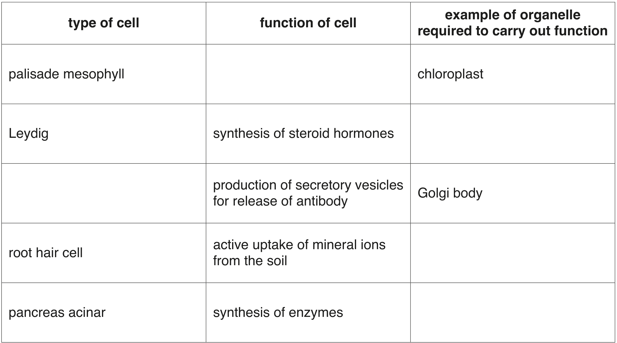
EduNinja

A-Level CAIE Biology AS1.2 Cells as Basic Units of Living OrganismsQuestion Bank

Question 1

[Maximum number: 9]

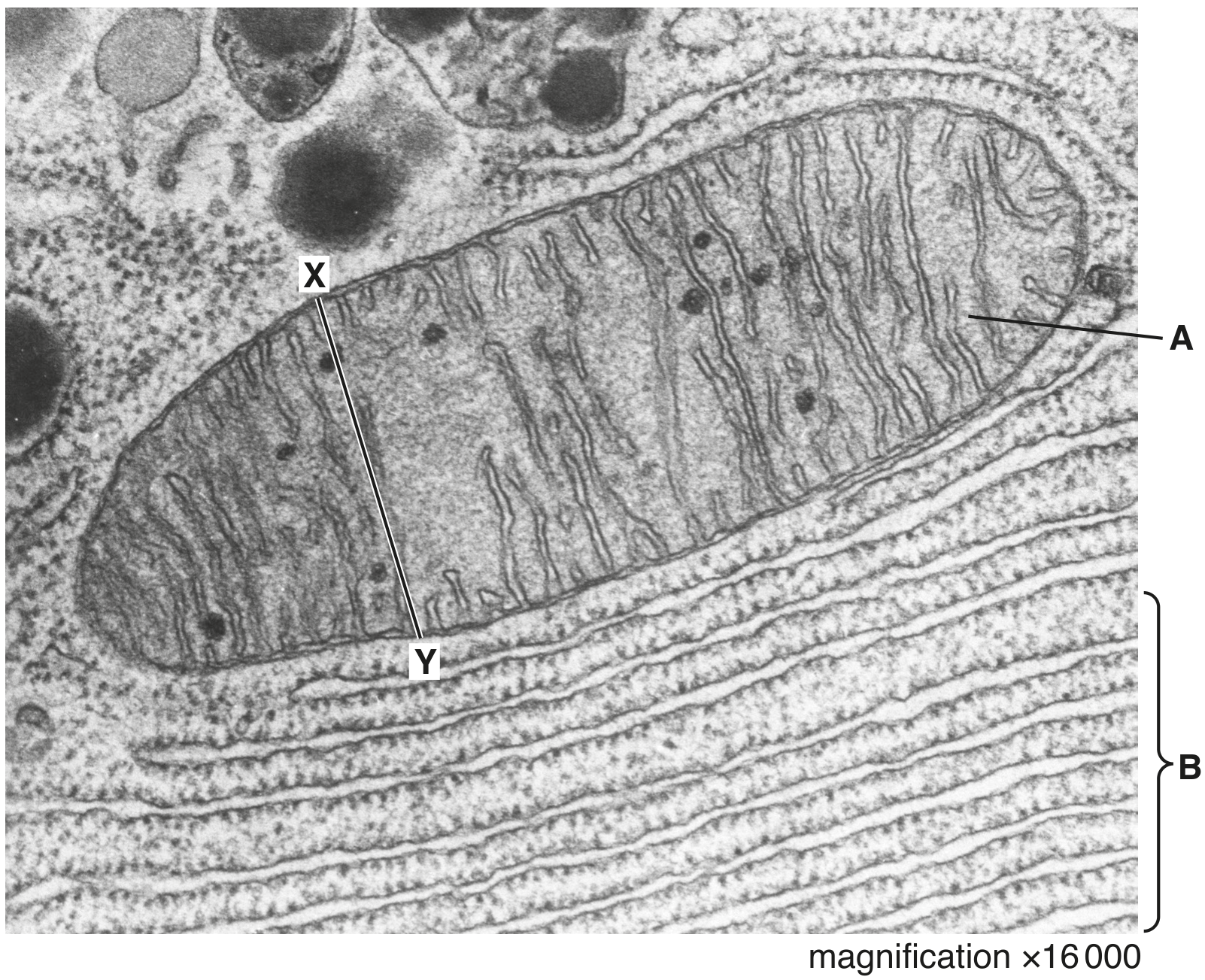
Fig. 1.1 is a transmission electron micrograph of a cell from the root of thale cress, Arabidopsis thaliana.
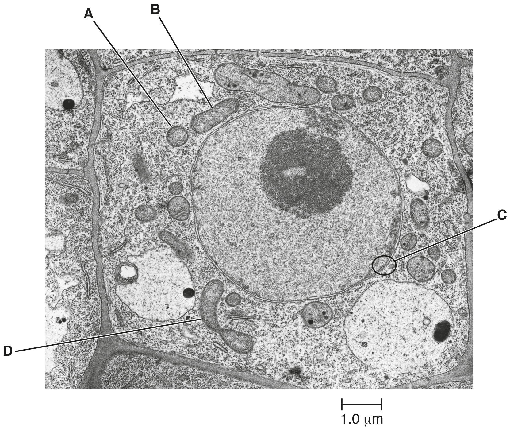
Fig. 1.1

Fig. 1.1

Question 1(a)

Question 1(a)(i)

(a)
(i)

The structures labelled A and B on Fig. 1.1 are sections of two mitochondria.

Suggest why A and B are different shapes.

[ 1 ]

Question 1(a)(ii)

(ii)

The structure labelled D on Fig. 1.1 is a mitochondrion about to divide.

Explain the importance of the division of mitochondria for the cell shown in Fig. 1.1 and for cells in the root tips of thale cress.

[ 2 ]

Question 1(b)

(b)

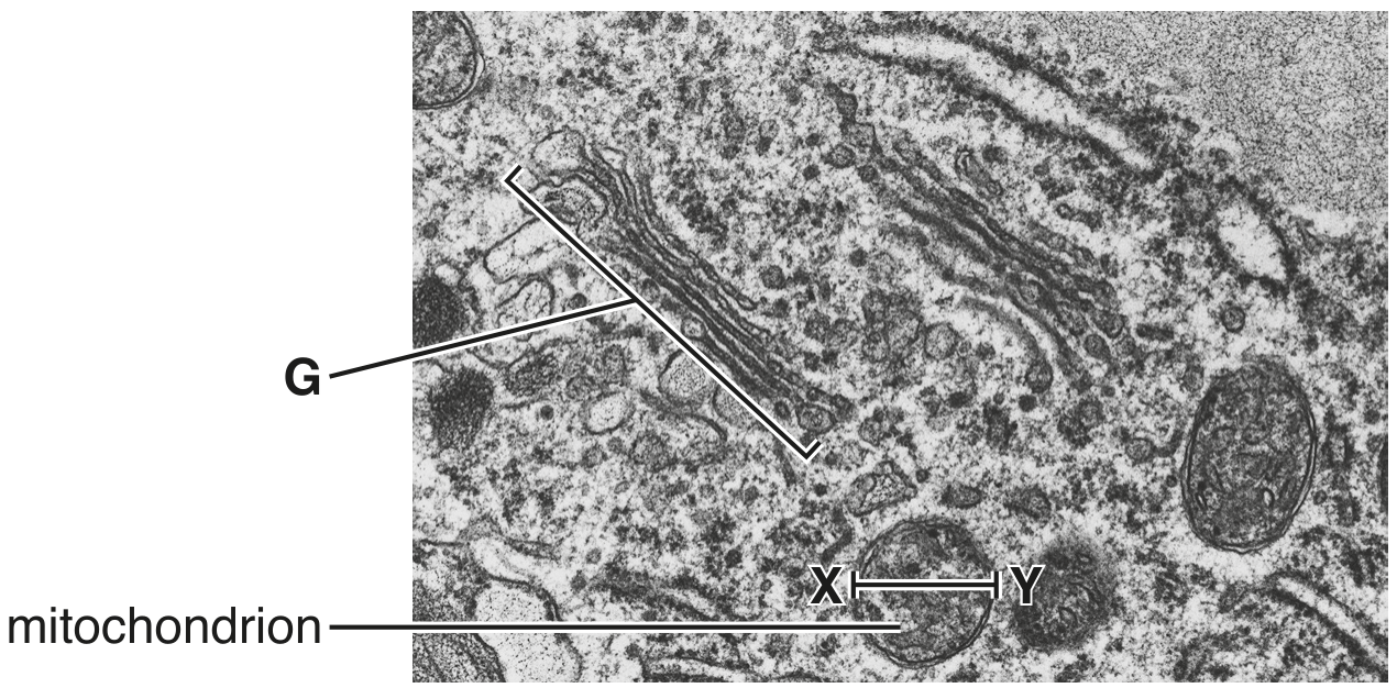
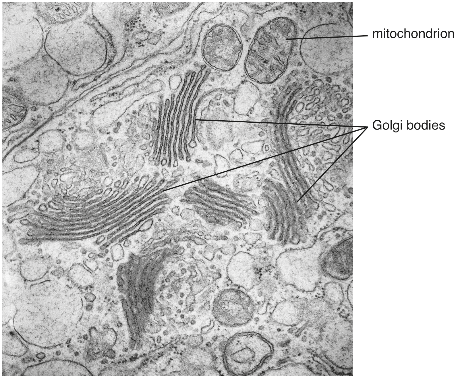
Within a cell, substances move between the nucleus and the cytoplasm. The area labelled C in Fig. 1.1 shows an area where this communication occurs.

Make a large, labelled drawing of area C to show where this communication occurs.

[ 2 ]

Question 1(c)

(c)

Outline the functions of the nucleus in non-dividing cells, such as the cell in Fig. 1.1.

[ 4 ]

Question 1

Question 1(a)

(a)

Each of the statements A to D describes a structure found in eukaryotic cells.
Identify the structure that is described in each statement.

A

An organelle that contains 70 S ribosomes.

B

A thread-like structure composed of DNA and histone proteins.

C

The organelle that modifies and packages proteins for secretion.

D

The structure that synthesises rRNA and combines it with proteins.

[ 4 ]

Question 1(b)

(b)

Prokaryotes and plant cells have cell walls.

Outline the composition of the cell wall of a prokaryote and the composition of the cell wall of a plant cell to show how they differ.

[ 2 ]

Question 1

Question 1(a)

(a)

In multicellular organisms, the structure of different cell types is adapted to their function. Within these cells there are a number of different organelles, each with a particular function.

Table 1.1 contains information about the structure and function of five different types of cell. The table also includes, for each type of cell, one example of a cell organelle that is essential for the function to be carried out.

Complete Table 1.1.

Table 1.1

Table 1.1

[ 5 ]

Question 1(b)

(b)

A tissue is a collection of one or more types of cell, specialised to carry out a particular function.

An organ can be considered a structural unit within an organism that:
- consists of more than one type of tissue
- performs a particular function.

The aorta is the main artery of the body.
Explain, with reference to its structure and function, whether the aorta may be described as a tissue or an organ.

[ 4 ]

Question 1

[Maximum number: 1]

Which cell structure can be seen only with an electron microscope?

A

cell wall

B

chromosome

C

nucleolus

D

ribosome

Question 1

[Maximum number: 4]

Fig. 1.1 is a transmission electron micrograph of a part of an animal cell.

Fig. 1.1

Fig. 1.1

Question 1(b)

Question 1(b)(i)

(a)
(i)

Name the organelle A and state its role in cells.
name
role

[ 2 ]

Question 1(b)(ii)

(ii)

Name the cell structure labelled B and state one reason for your answer. name
reason

[ 2 ]

Question 1

[Maximum number: 2]

Fig. 1.1 is an electron micrograph of part of a eukaryotic cell.

Fig. 1.1

Fig. 1.1

×47000\times 47000

Question 1(b)

(a)

Both the Golgi body and the rough endoplasmic reticulum are part of the internal network of membranes in cells.

Outline structural features shown in Fig. 1.1 that identify G as the Golgi body and not the rough endoplasmic reticulum.

[ 2 ]

Question 1

Question 1(a)

(a)

Aphids are small insects which feed directly on phloem sap.
The salivary glands of aphids have secretory cells that make and release a variety of proteins that assist in feeding.

Fig. 1.1 is a transmission electron micrograph of a small area of a salivary gland cell of an aphid.

Fig. 1.1

Fig. 1.1

Describe the role of Golgi bodies in secretory cells, such as the salivary gland cells of aphids.

[ 3 ]

Question 1(b)

Question 1(b)(i)

(b)
(i)

Explain why secretory cells have large numbers of mitochondria.

[ 2 ]

Question 1(b)(ii)

(ii)

Mitochondria are partly controlled by the nucleus, but can also function independently.

Suggest the features of mitochondria that allow them to function independently of the nucleus.

[ 2 ]

Question 1(c)

(c)

Aphids are important vectors of plant viral diseases.

[ 4 ]

Question 1(c)(i)

(i)

Describe the structure of a typical virus.

[ 3 ]

Question 1(c)(ii)

(ii)

Suggest how viruses are able to pass from one plant cell to the next without crossing membranes.

[ 1 ]

Question 1

[Maximum number: 2]

Fig. 1.1 is a diagram of a molecule of haemoglobin.

Fig. 1.1

Fig. 1.1

Question 1(a)

Question 1(a)(i)

(a)
(i)

Name the structure labelled A on Fig. 1.1.

[ 1 ]

Question 1(a)(ii)

(ii)

State the function of structure A.

[ 1 ]

Question 1

[Maximum number: 5]

Fig. 1.1 is a photomicrograph of a transverse section through the stem of creeping buttercup, Ranunculus repens.

Fig. 1.1

Fig. 1.1

Question 1(d)

(a)

Draw a diagram in the space provided to show the structure of a typical plant cell.

Label your diagram with the name of any structures that are found only in plant cells.
Do not label any structures that are also found in animal cells.

[ 5 ]

Question 1

[Maximum number: 5]

Match the description for each of statements A to E to a correct cell structure.

A Double membrane-bound organelle, absent in animal cells, that produces ATP.

B Partially permeable membrane surrounding the large permanent vacuole of plant cells.

C Formed from microtubules during mitosis.

D Has peptidoglycan as one of its major components. E Site of assembly of 80 S ribosomes.

0 selected