Question 1
Question 1(c)
(a)
State two components of a cell surface membrane other than phospholipid molecules and describe their function.
component 1
function
component 2
function
[ 4 ]
 EduNinja
State two components of a cell surface membrane other than phospholipid molecules and describe their function.
component 1
function
component 2
function
Fig. 1.1 represents part of the wall of a proximal convoluted tubule (pct) in a kidney nephron.
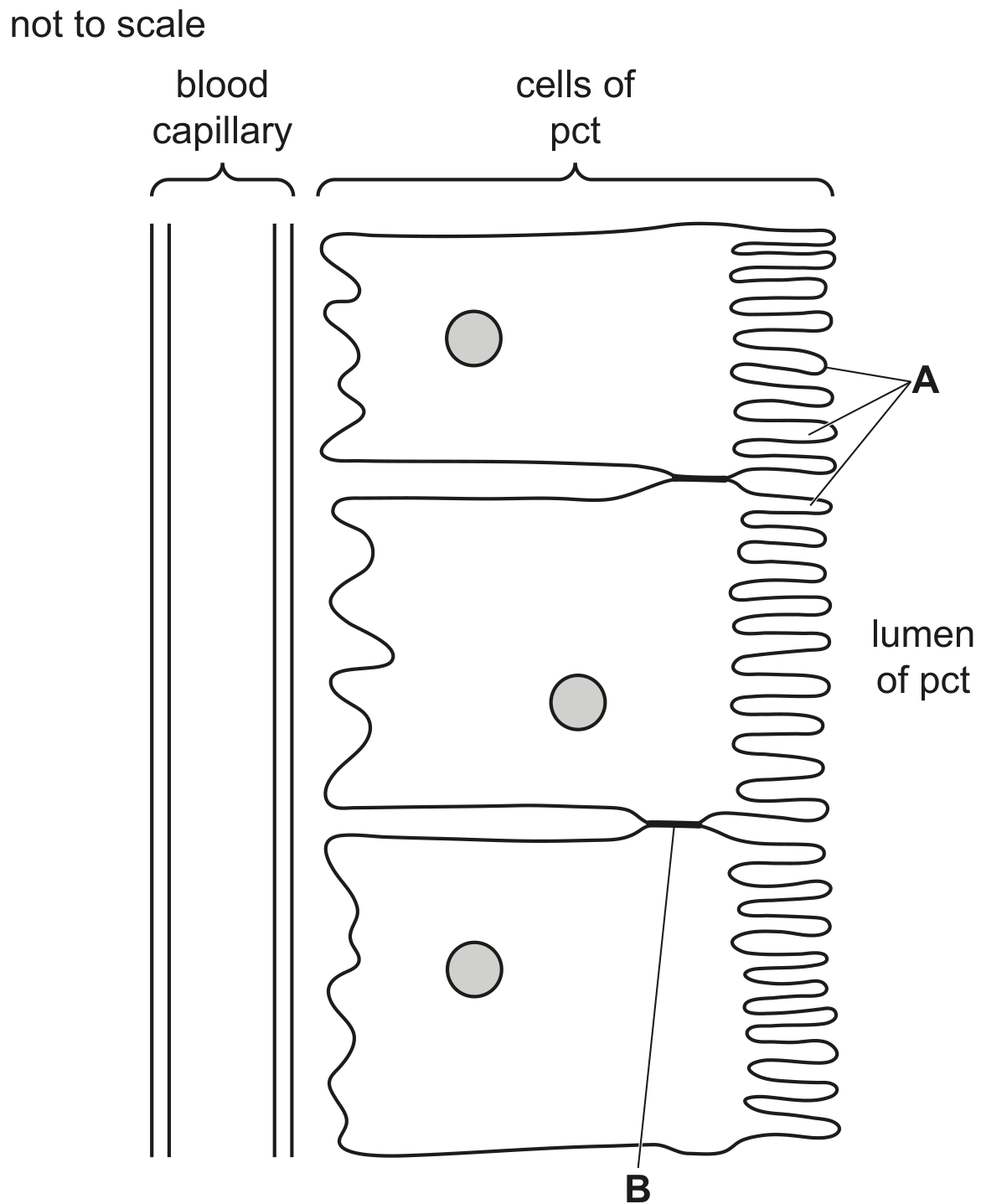
Fig. 1.1
Name the features of the wall of a pct that are labelled A and B in Fig. 1.1.
A
B
Fig. 1.1 shows a diagram of part of a cell surface membrane.
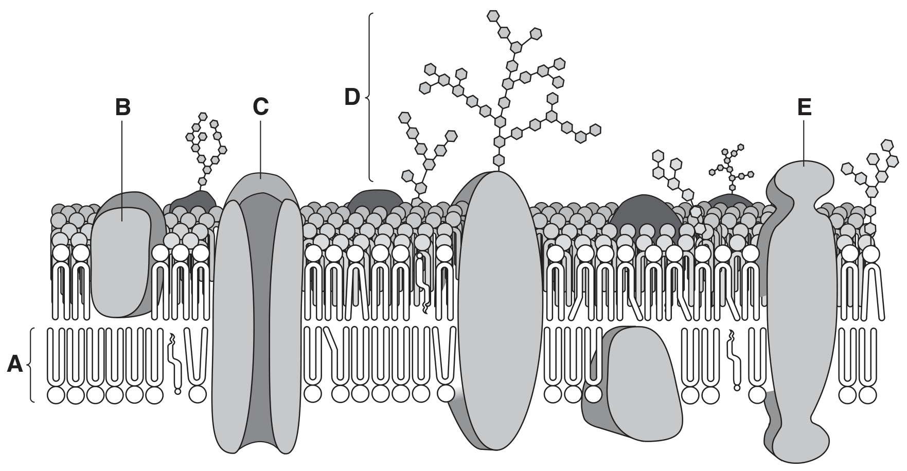
Fig. 1.1
Explain how the features of molecules of A cause them to form a layer in the membrane as seen in Fig. 1.1.
State the functions of C and D.
C
D
Use a label line and the appropriate letter to label each of the following on Fig. 1.1.
P protein for active uptake of potassium ions
Q protein for facilitated diffusion of polar molecules
R receptor site for a hormone
S hydrophilic heads of phospholipids on the internal surface of the membrane
T molecule that modifies the fluidity of the membrane
A diagram of a chromosome from a dividing cell is shown in Fig. 1.1.

Fig. 1.1
The control of the cell cycle can be affected by extracellular chemical messengers that bind to proteins and glycoproteins in the cell surface membrane. The overall mechanism is known as cell signalling.
State the term used to describe the proteins and glycoproteins that function in this way.
Fig. 1.1 is a diagram showing part of a cell surface membrane of an animal cell.
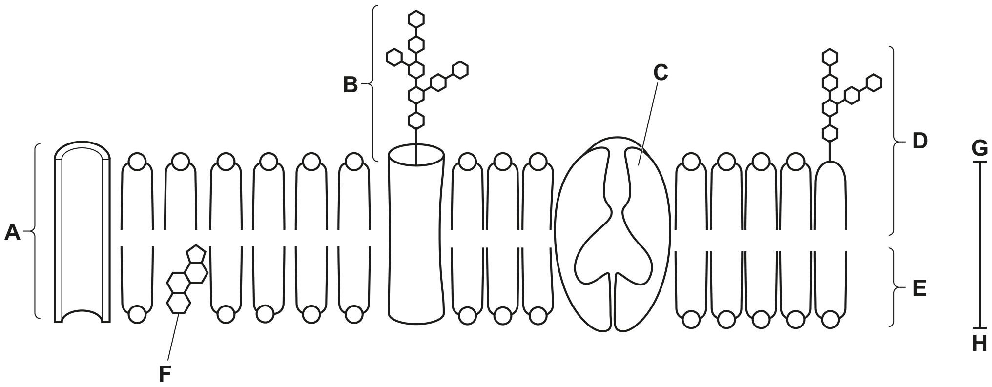
Fig. 1.1
State the approximate thickness of the membrane as shown by the line G-H.
Complete Table 1.1 to show:
- the names and functions of the components of the cell surface membrane
- the letters of the labels in Fig. 1.1 that identify each component.
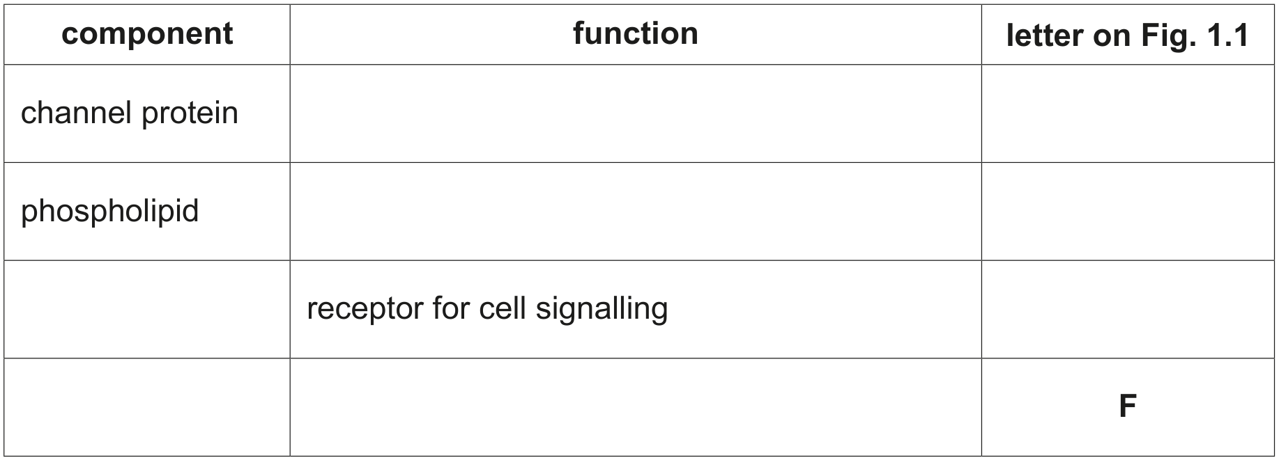
Table 1.1
The cell surface membrane has a fluid mosaic structure.
Describe what is meant by the term fluid mosaic.
In 1934, the biologists Davson and Danielli published their suggestion for the structure of the cell surface membrane, as shown in Fig. 1.1.
They suggested that the membrane was a phospholipid bilayer with a layer of hydrophilic protein on both surfaces.
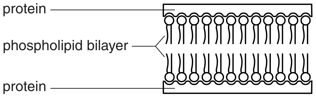
Fig 1.1
State one way in which the Davson-Danielli structure is similar to the fluid mosaic structure and one way in which it differs from the fluid mosaic model.
similarity
difference
Fig. 1.1 is a diagram of a transverse section through a vein.
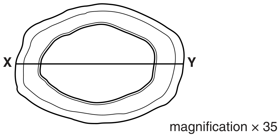
Fig. 1.1
Explain how the following structural features of a capillary are related to its function.
The capillary wall is composed of a single layer of squamous epithelial cells.
Fig. 1.1 is an electron micrograph of part of a eukaryotic cell.
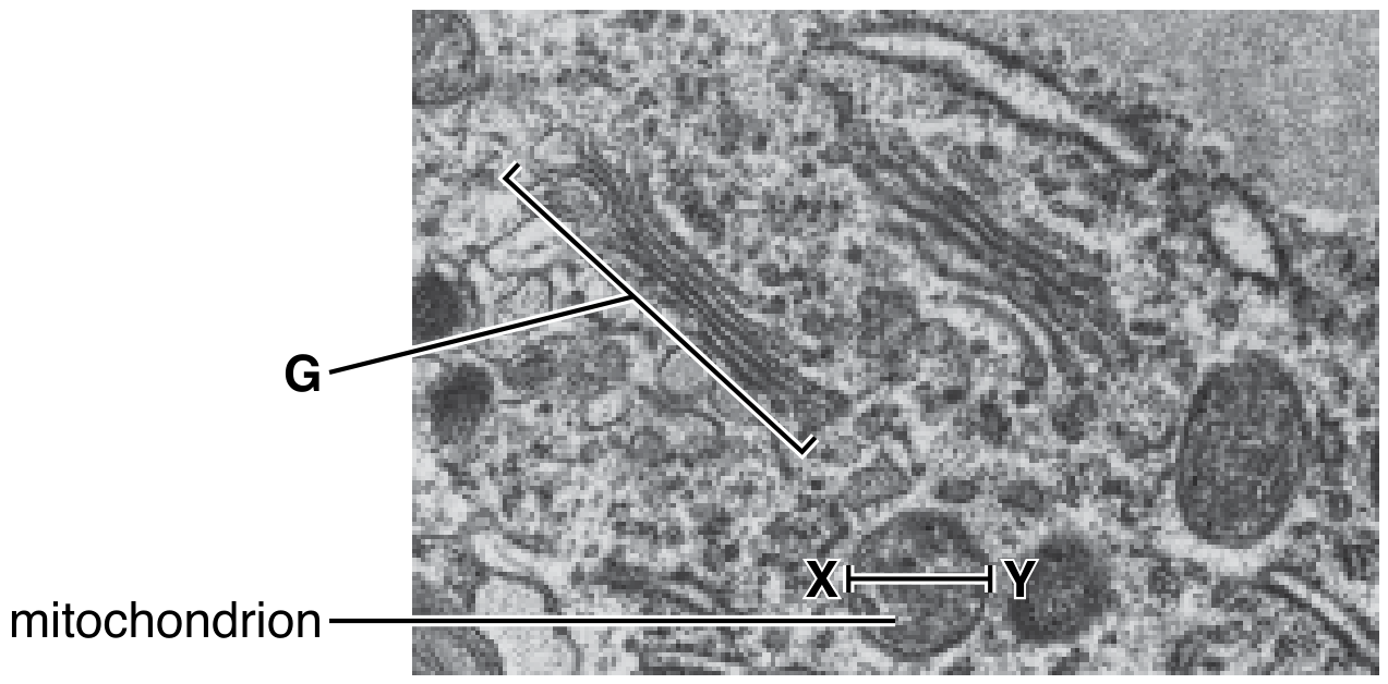
Fig. 1.1
×47000
Many of the cell structures in Fig. 1.1 are surrounded by membranes.
Membranes are approximately 6 nm to 7 nm wide.
Describe the fluid mosaic model of membrane structure.
There is space below for a diagram.
The inner membrane of the mitochondrial envelope is much less permeable than the outer membrane.
Suggest one way in which the structure of the inner membrane of the mitochondrion may differ from that of the outer membrane to produce a less permeable inner membrane.
Fig. 1.1 is an electron micrograph of a transverse section of palisade mesophyll tissue in the leaf of the flowering plant, Zinnia elegans.
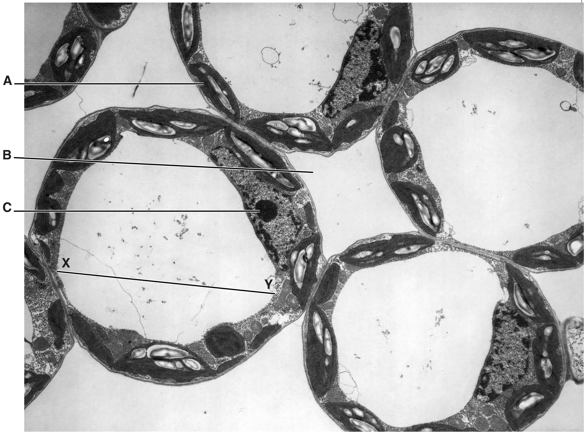
Fig. 1.1
magnification
The membrane surrounding the vacuole, called the tonoplast, has a fluid mosaic structure.
Describe the structure of this membrane.