C2.2 Neural signalling

Neural signalling transmits information through neuron structure, ion gradients, action potentials, synapses and chemical modulation of postsynaptic responses, linking cellular mechanisms to rapid communication in nervous systems.

Syllabus
First assessment 2025
Topic
C2.2
Level
SL

Learning objectives

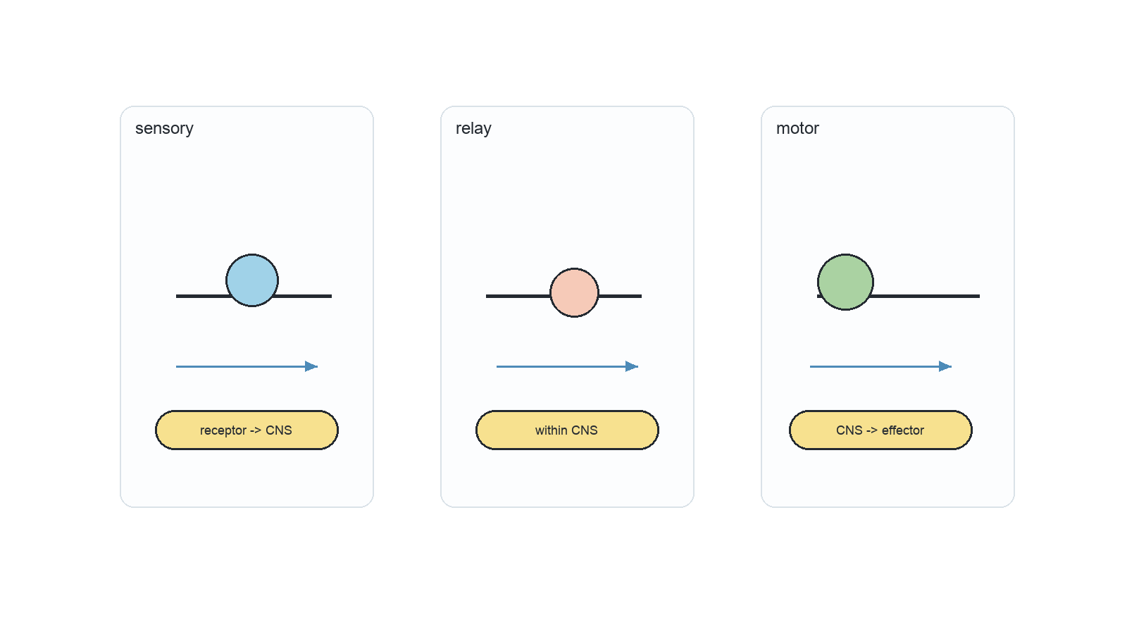
Neuron Shape Matches the Direction of Information Flow

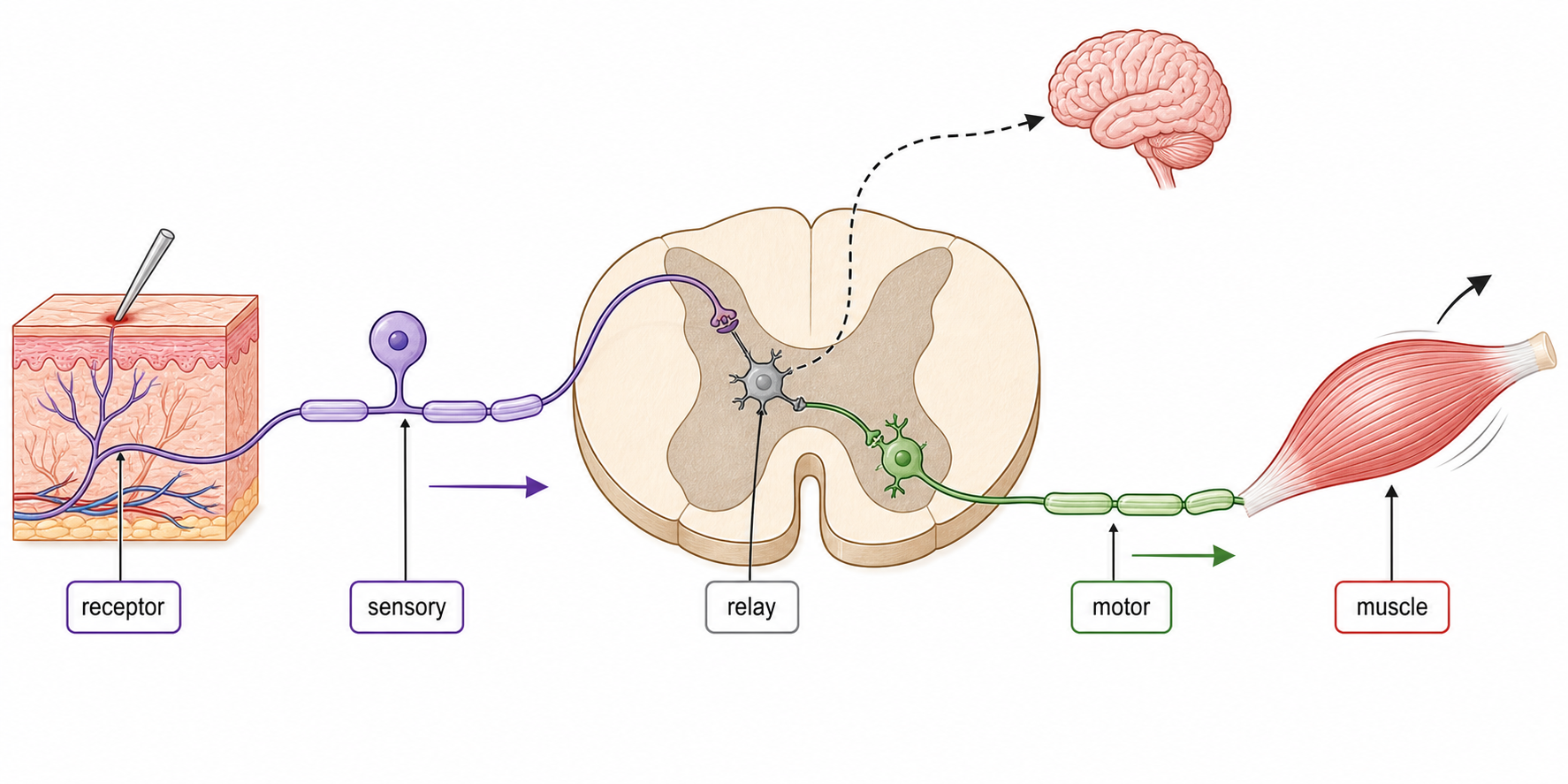
Neuron Information route Decisive structural pattern
sensory receptor → CNS long dendron toward an off-centre cell body; axon continues into CNS
relay within CNS many short dendrites and a short axon connect neurons locally
motor CNS → muscle or gland many dendrites around the cell body; one long axon reaches an effector
Sensory, relay and motor neurons compared by cell-body position, fibre arrangement and direction of impulse flow.

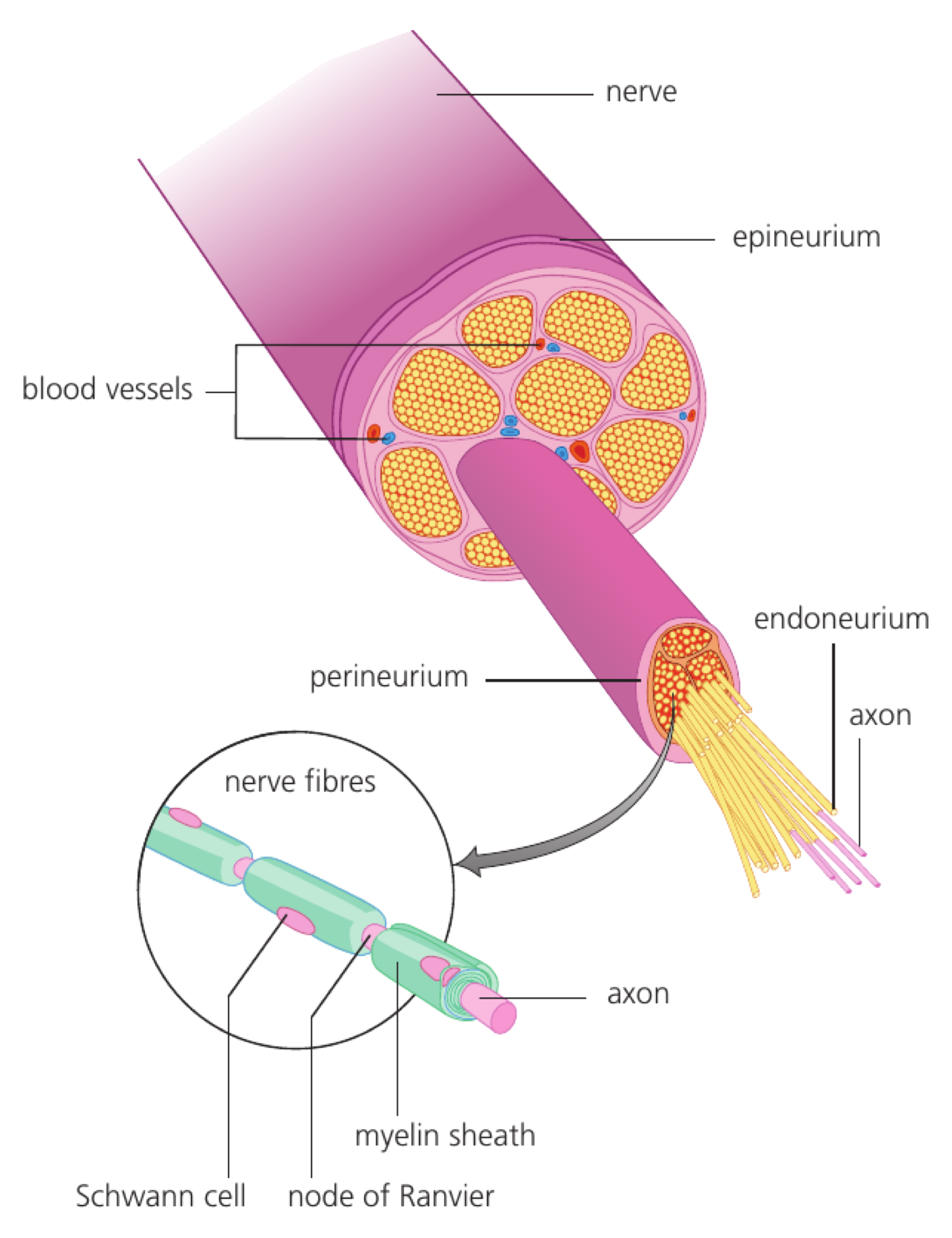
Dendrites and dendrons carry impulses toward the cell body; an axon carries impulses away from it. A nerve is a bundle of many nerve fibres, not one neuron.

Resting Potential Stores Energy in Unequal Ion Distributions

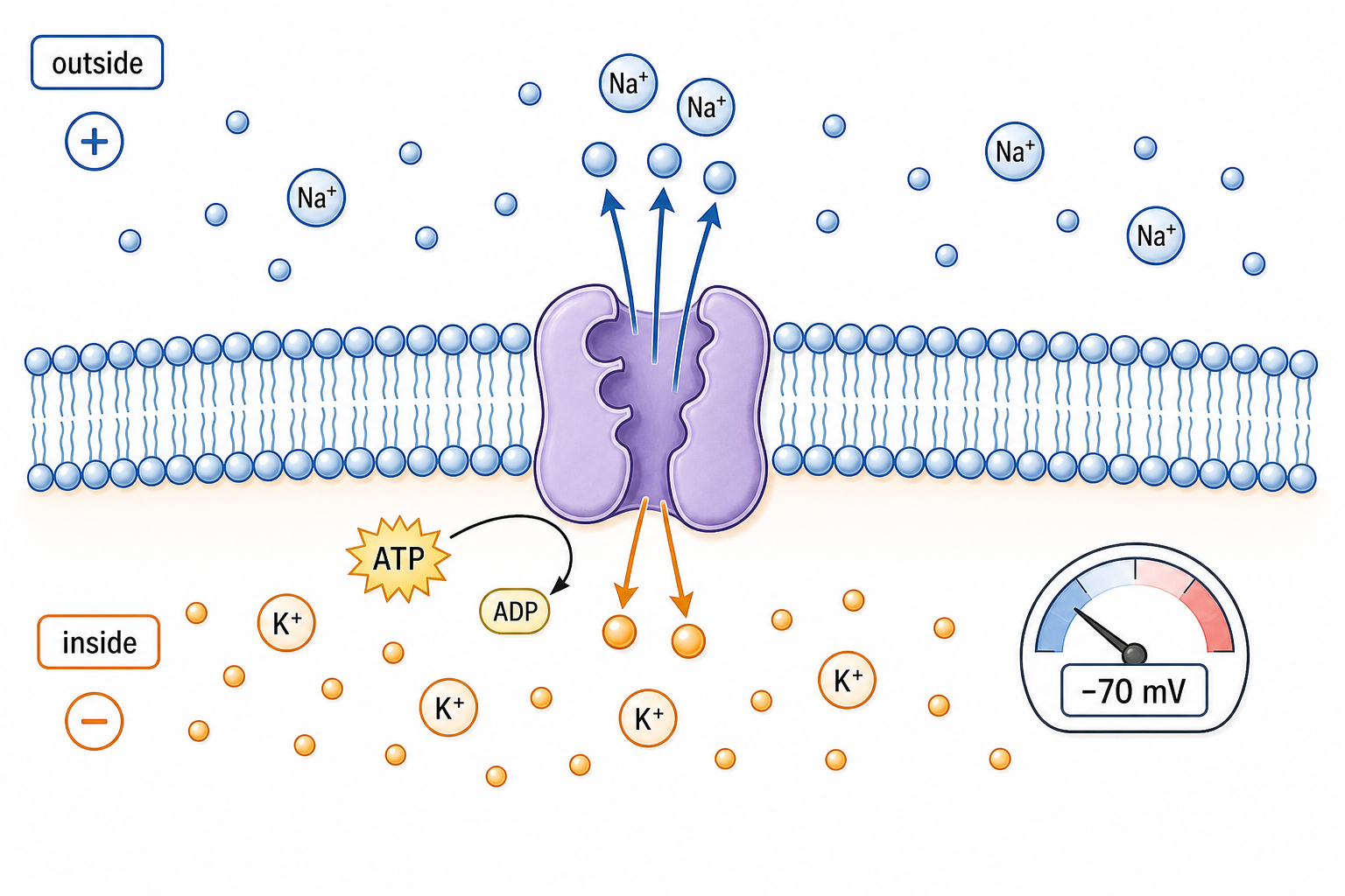
At rest, an axon's inside is about −70 mV relative to the outside. The membrane is polarized because ions and large negative solutes are distributed unequally across it.

The sodium–potassium pump hydrolyses ATP to move 3 Na⁺ out for every 2 K⁺ in, against both concentration gradients. It establishes and maintains high Na⁺ outside and high K⁺ inside.

At rest the membrane is much more permeable to K⁺ than to Na⁺. K⁺ leakage leaves behind impermeant negative charge, so the inside becomes negative. The pump maintains the gradients that this leakage continually uses.

A resting axon is shown with a negative interior relative to the positively charged exterior.

A Nerve Impulse Is a Regenerated Action Potential

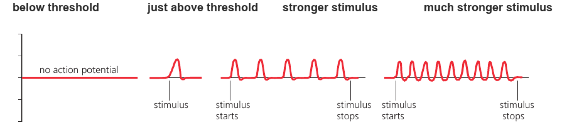
An action potential is a brief reversal of membrane potential, typically from about −70 mV to about +40 mV. A nerve impulse is this action potential repeatedly regenerated along a nerve fibre.

  • Below threshold, the membrane returns to rest without firing.
  • At or above threshold, a full action potential occurs.
  • Stronger stimuli do not make larger action potentials; they produce a higher frequency of standard-sized impulses.
Traces show no action potential below threshold, one standard-sized action potential just above threshold, and higher impulse frequency with stronger sustained stimuli.

Ion flow through open channels is passive, down electrochemical gradients. ATP is used by pumps to maintain and restore those gradients, not to push each ion through an open channel.

Axon Design Sets the Speed of an Impulse

Structural change Immediate physical effect Consequence for speed
larger axon diameter lower resistance to local current inside the axon adjacent membrane reaches threshold sooner
myelin sheath raises membrane resistance and limits ion exchange to nodes of Ranvier local current travels farther before another action potential is generated

Within both myelinated and unmyelinated groups, conduction velocity increases with diameter. At the same diameter, a myelinated axon is much faster; giant unmyelinated axons partly compensate through size.

Diameter and myelination are independent adaptations. Neither changes the amplitude of each action potential; they change how quickly the next region reaches threshold.

A Chemical Synapse Has a Sender and a Receiver

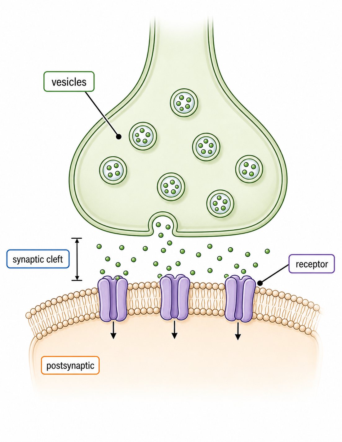
  • The presynaptic terminal stores neurotransmitter in vesicles and releases it.
  • The synaptic cleft is a narrow extracellular gap, typically about 20 nm.
  • The postsynaptic membrane carries receptors that convert the chemical signal into a membrane response.

Transmission is normally one-way because release machinery is presynaptic while matching receptors are postsynaptic. Synapses can connect neuron to neuron, neuron to muscle, or neuron to a gland cell.

A presynaptic terminal contains neurotransmitter vesicles above a narrow synaptic cleft and a postsynaptic membrane bearing receptor proteins.

Calcium Converts an Arriving Impulse into Secretion

  1. An action potential depolarizes the presynaptic terminal.
  2. Voltage-gated Ca²⁺ channels open.
  3. Ca²⁺ diffuses into the terminal down its electrochemical gradient.
  4. Ca²⁺ triggers neurotransmitter vesicles to dock and fuse with the presynaptic membrane.
  5. Exocytosis releases neurotransmitter, creating a high concentration beside the cleft.

This is an electrical → chemical conversion. Ca²⁺ acts as an intracellular signal that couples membrane depolarization to vesicle fusion.

Presynaptic structure Contribution
mitochondria supply ATP for vesicle cycling and ion-gradient maintenance
Golgi apparatus and vesicles process, package and store transmitter components
presynaptic membrane provides voltage-gated Ca²⁺ channels and the fusion surface

An EPSP Moves the Postsynaptic Membrane toward Threshold

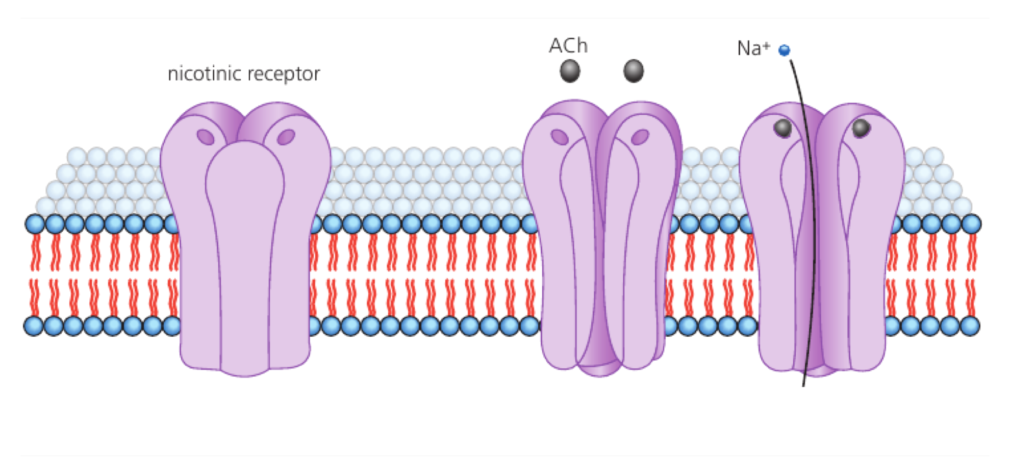
neurotransmitter diffuses across the cleft → binds a complementary postsynaptic receptor → ligand-gated cation channels open → Na⁺ entry depolarizes the membrane → an excitatory postsynaptic potential (EPSP) forms

An EPSP is graded: more transmitter can produce a larger local depolarization. It makes an action potential more likely, but only a summed depolarization reaching threshold triggers the all-or-none spike.

Acetylcholine binding opens a postsynaptic nicotinic receptor channel and allows sodium ions to enter.

The signal must be removed. At cholinergic synapses, acetylcholinesterase hydrolyses acetylcholine; receptors close, and choline can be taken up for reuse. Other transmitters may be removed by reuptake or diffusion.

SL Summary: Trace the Signal into the Next Cell

Within a neuron: ion gradients create a resting potential; threshold opens a regenerative membrane event; axon diameter and myelin change how quickly it travels.
Across a synapse: arriving depolarization opens Ca²⁺ channels → Ca²⁺ triggers exocytosis → transmitter diffuses → postsynaptic receptors open channels → an EPSP moves the next cell toward threshold.

If transmission fails here… Inspect…
resting voltage cannot be maintained ATP supply, Na⁺/K⁺ pump and membrane permeability
presynaptic impulse arrives but no transmitter appears voltage-gated Ca²⁺ channels and vesicle fusion
transmitter reaches the cleft but no EPSP forms receptor match, postsynaptic channel and transmitter removal

Neurons exam focus

5 marks

Draw a labelled diagram of a motor neuron.

Resting potential generation

4 marks

Outline how neurons generate a resting potential.

Nerve impulses as action potentials

3 marks

Outline how nerve impulses are transmitted along a nerve fibre.

Variation in impulse speed

1 mark

The diagrams represent sections through different axons. Which axon has the slowest speed of impulse?
Synapses as junctions

2 marks

Distinguish between the presynaptic and postsynaptic membranes by giving two differences.
Neurotransmitter release

3 marks

Describe how neurotransmitters are released from a presynaptic neuron membrane.

Excitatory postsynaptic potential

3 marks

Describe how an excitatory postsynaptic potential is generated.

Up to one additional mark is available for the construction of your answer.