D3.1.4—Human reproductive system anatomy

Human reproductive system anatomy identifies ovaries, oviducts, uterus, cervix, vagina, testes, epididymis, ducts, glands and penis by structure and function.

Syllabus
First assessment 2025
Objective
D3.1.4
Level
SL

Exam analysis

Chance of appearing7%of analysed past papers
Latest appearanceMay 2025
Most common paperPaper1
Typical marks1

Common command terms

  • Identify
  • Draw

Scoring notes

Common mistake
Misidentifying epididymis, sperm duct, prostate and seminal vesicles on male diagrams.

Recent exam appearances

May 2025Paper1A ["SL"] · TZ119[ 1 ]D3.1.4—Human reproductive system anatomy
May 2021Paper1 ["SL"] · TZ230[ 1 ]D3.1.4—Human reproductive system anatomy
May 2019Paper1 ["SL"] · TZ130[ 1 ]D3.1.4—Human reproductive system anatomy
May 2017Paper1 ["SL"] · TZ128[ 1 ]D3.1.4—Human reproductive system anatomy
May 2015Paper2 ["SL"] · TZ16(a)[ 5 ]D3.1.4—Human reproductive system anatomy

Coverage 2011–2025 · Updated 16 Jul 2026

The Male Reproductive System Produces, Matures and Delivers Sperm

seminiferous tubules in testes → epididymis → sperm duct → fluids added by seminal vesicles and prostate → urethra → penis

Structure Essential function
testes in scrotum make sperm and testosterone at a temperature below body core
epididymis completes sperm maturation and stores sperm
sperm duct transports sperm during ejaculation
seminal vesicles and prostate add nutrient-containing, alkaline seminal fluid
urethra and penis conduct and deliver semen outside the body

Semen is sperm plus glandular fluid. It is not produced entirely in the testes, and semen production is not the same process as spermatogenesis.

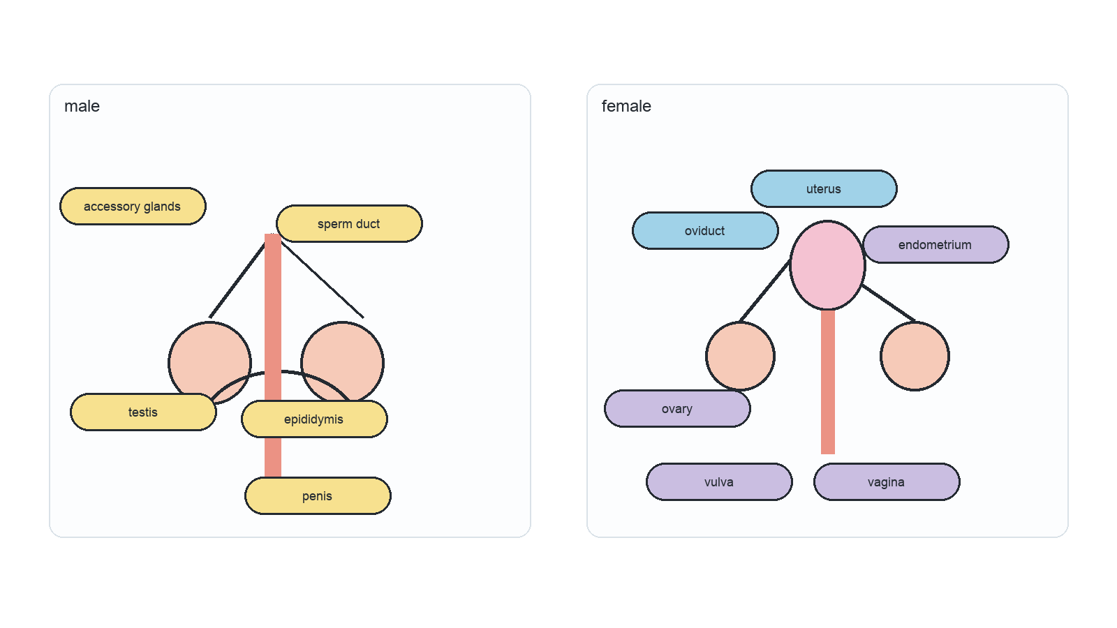
Paired male and female human reproductive-system diagrams label the main organs and connecting ducts.

The Female Reproductive System Connects Ovulation, Fertilization, Implantation and Birth

ovary releases secondary oocyte → oviduct receives and transports it; fertilization usually occurs in the upper oviduct → embryo reaches uterus → implantation occurs in endometrium → cervix and vagina form the exit route at birth

Structure Essential function
ovary produces oocytes, oestradiol and progesterone
oviduct transports oocyte or embryo; usual fertilization site
uterus muscular organ supporting embryo and foetus
endometrium vascular lining for implantation and early support
cervix muscular neck between uterus and vagina
vagina and vulva canal and external structures for intercourse and birth

Draw the oviduct opening into the uterine cavity, not ending inside the uterine wall. The ovary lies near the funnel-shaped end but is not directly joined by a sealed tube.

Human Reproduction Coordinates Ploidy, Place and Hormone Timing

meiosis halves chromosome-set number and reshuffles alleles → specialized haploid gametes form → random fertilization restores diploidy → mitosis grows the genetically unique zygote

Transition Main location Key control or structure
sperm production and maturation testis → epididymis FSH/testosterone; seminiferous tubules
follicle growth and ovulation ovary FSH, oestradiol and LH surge
fertilization upper oviduct gamete encounter and membrane fusion
implantation preparation endometrium oestradiol and progesterone
IVF intervention clinic/laboratory → uterus controlled stimulation, retrieval, culture and transfer

When explaining a reproductive event, name the chromosome state, the anatomical location and the controlling signal. Mixing these three levels is the main source of fragmented answers.

Human reproductive system anatomy

[ 6 ]

Draw a labeled diagram of the female reproductive system.

Drawing canvas

Concept essentials

  • The epididymis is where sperm complete maturation and become motile.
  • The oviduct is the usual site of fertilization in humans.
  • Ovaries and testes both produce gametes and secrete hormones.