EduNinja

A-Level CAIE Biology A214.1 Homeostasis in MammalsQuestion Bank

Question 1

Question 1(a)

(a)

The water potential of mammalian blood needs to be maintained within narrow limits so that cells function efficiently. This process is called osmoregulation.

The relative medullary thickness (RMT) indicates the proportion of a kidney that is composed of medullary tissue.

 RMT = thickness of medulla  kidney size ×10\text { RMT }=\frac{\text { thickness of medulla }}{\text { kidney size }} \times 10

Table 1.1 shows the relationship between the RMT and the concentration of urine produced by four mammals from different habitats.
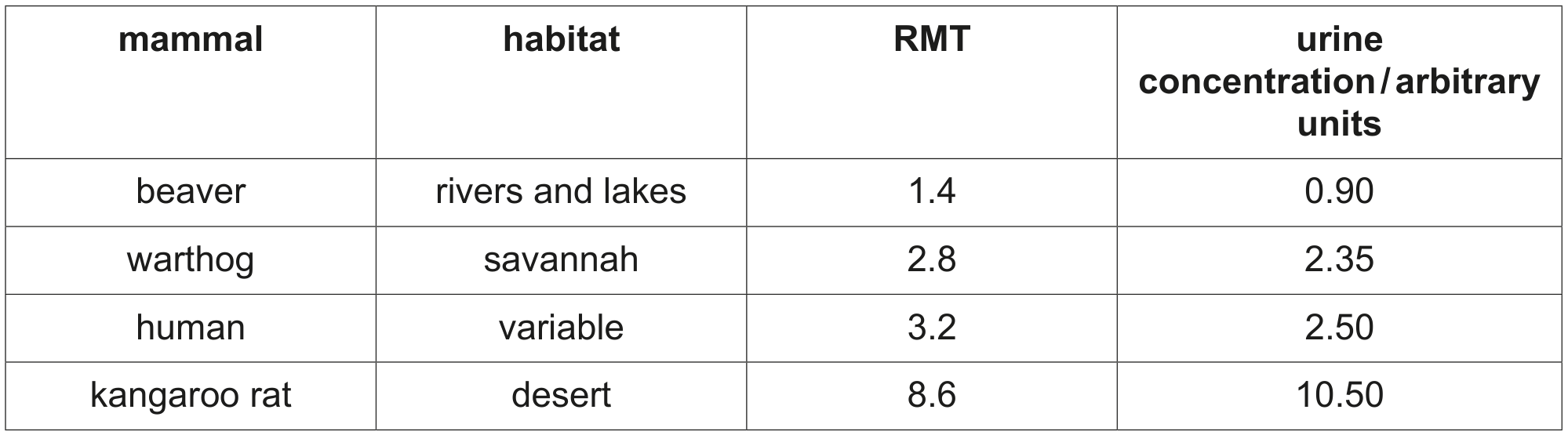
Table 1.1

Table 1.1

[ 3 ]

Question 1(a)(i)

(i)

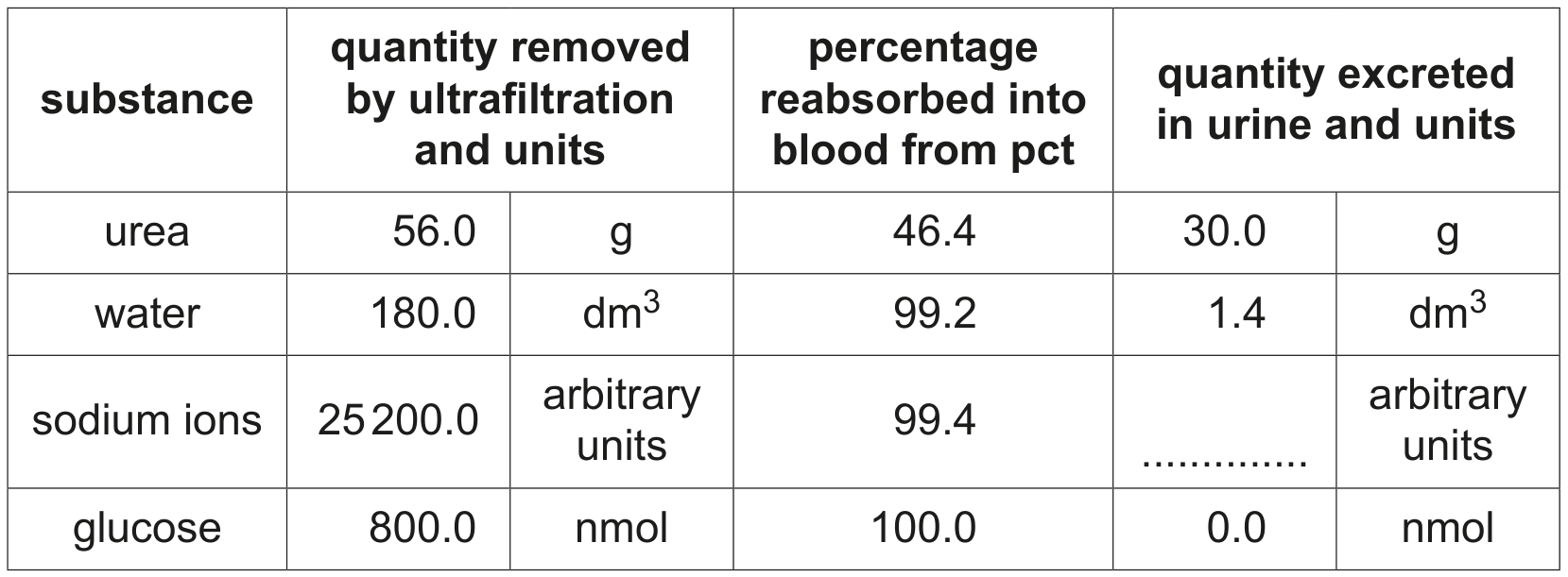
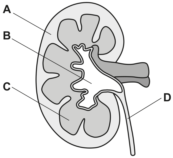
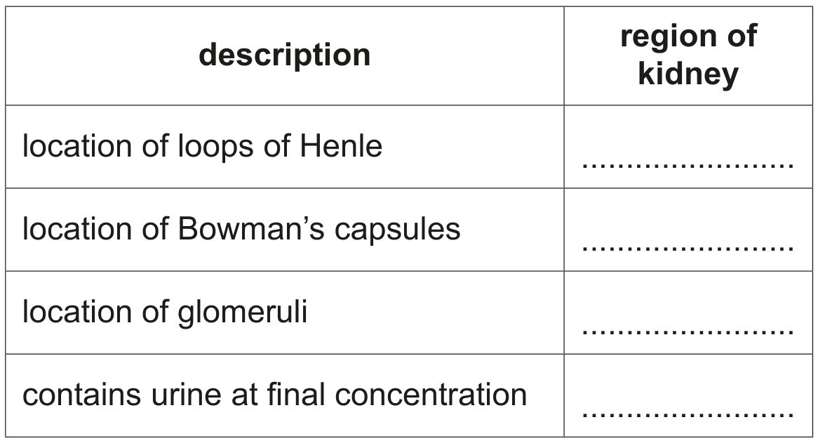
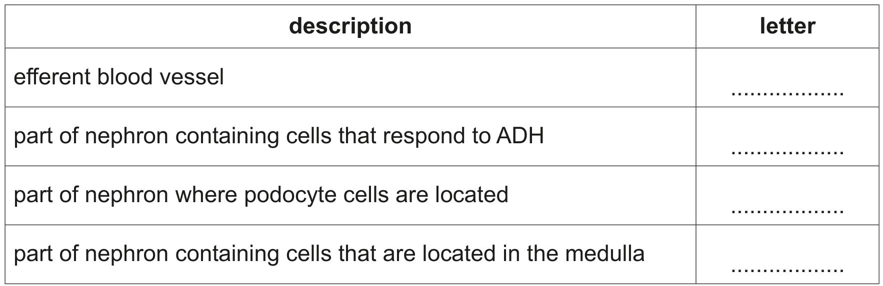
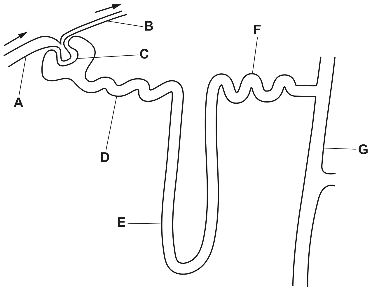
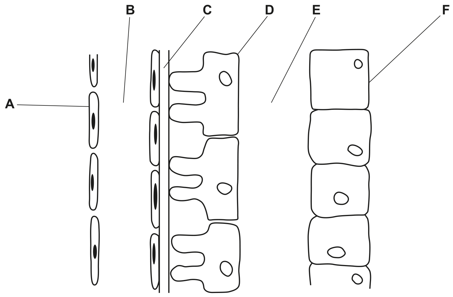
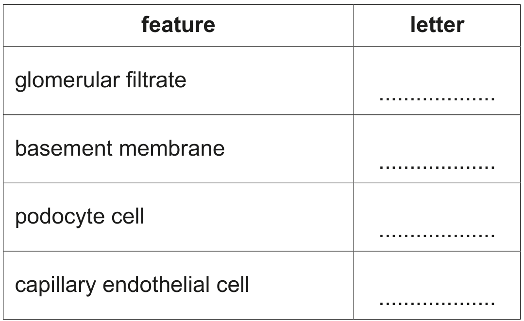
Name the parts of the nephron that are located in the medulla.

[ 2 ]

Question 1(a)(ii)

(ii)

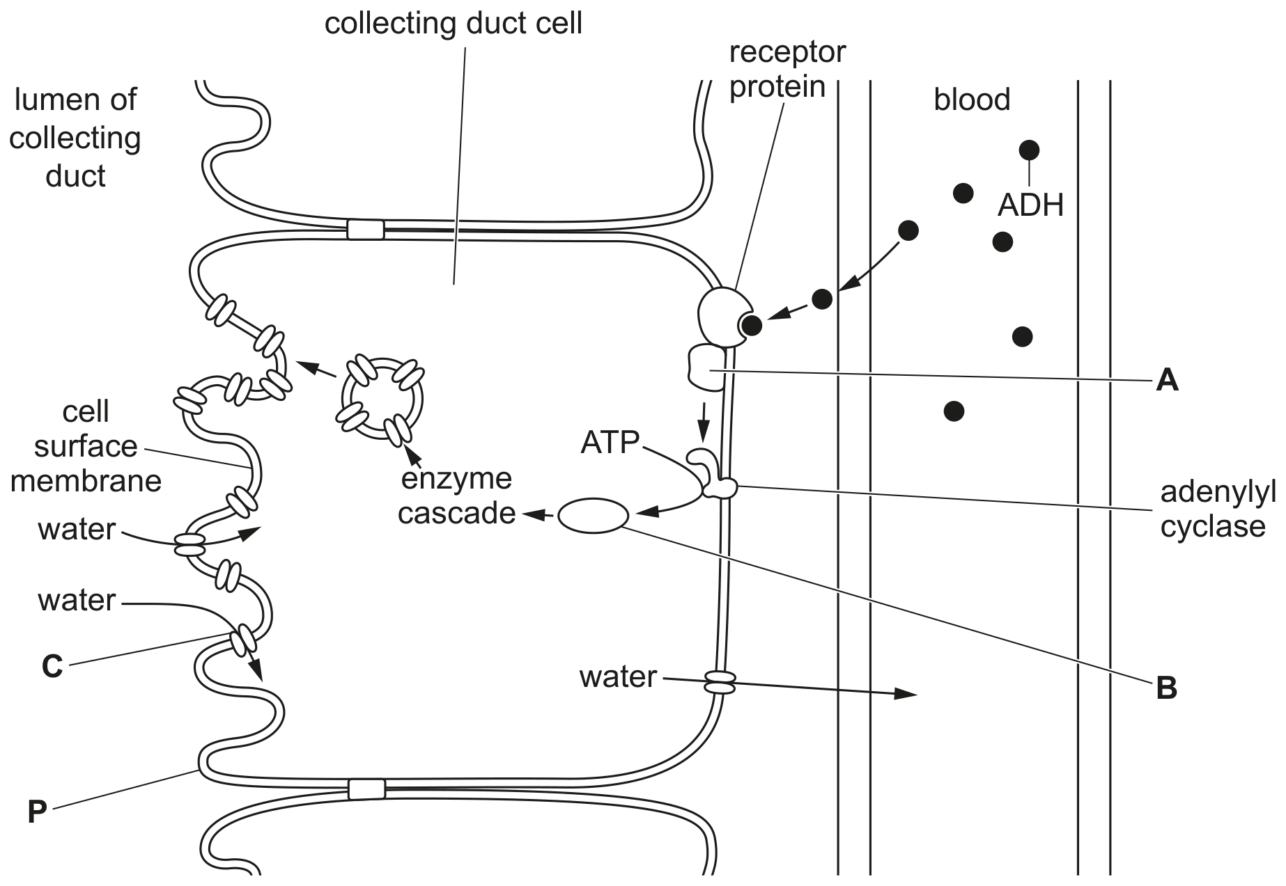
Name a hormone involved in osmoregulation.

[ 1 ]

Question 1(b)

(b)

The warthog, Phacochoerus africanus, is a member of the pig family. The warthog lives in dry savannah areas of sub-Saharan Africa.

Fig. 1.1 shows a warthog.
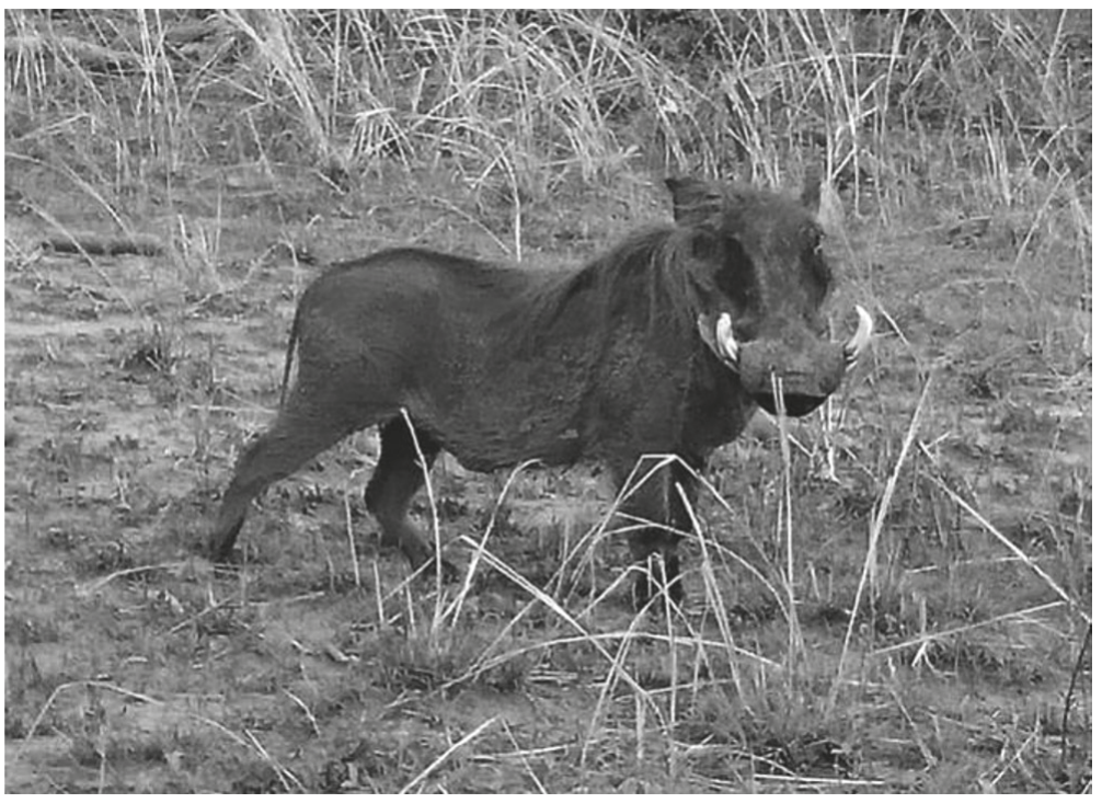
Fig. 1.1

Fig. 1.1

A warthog and a human have similar values of RMT and concentration of urine. A human can survive only a few days without drinking water, whereas a warthog can live for several months without drinking water.

Suggest how a warthog is able to survive several months without drinking water.

[ 2 ]

Question 1

Question 1(b)

(a)

Table 1.1 shows the quantities, per day, of some of the substances that are:
- removed from the blood by ultrafiltration
- reabsorbed into the blood by the pct
- excreted in the urine.

Table 1.1

Table 1.1

Complete Table 1.1 by calculating the quantity of sodium ions excreted in the urine.
Write your answer in the table to one decimal place.
Show your working in the space below.

[ 2 ]

Question 1

Question 1(a)

(a)

In mammals, the hormone ACTH is released from the anterior pituitary gland. ACTH stimulates the cells of the adrenal glands to synthesise the hormone cortisol. This is an example of cell signalling.

The main stages of cell signalling involving ACTH are similar to the main stages of cell signalling by adrenaline.

Describe the main stages of cell signalling involving ACTH.

[ 5 ]

Question 1(b)

(b)

If the concentration of cortisol in the blood increases above its set point, it will inhibit the production of ACTH by the anterior pituitary gland.

Name this type of homeostatic mechanism.

[ 1 ]

Question 1(d)

(c)

One symptom of Cushing's disease in a dog is that the dog will want to drink much more than normal.

Suggest one other reason why a dog would want to drink much more than normal.

[ 1 ]

Question 1

Question 1(a)

(a)

Fig. 1.1 is a drawing of a longitudinal section (LS) of a human kidney.

Fig. 1.1

Fig. 1.1

Use the letters A, B, C and D in Fig. 1.1 to complete Table 1.1.
Each letter may be used once, more than once or not at all.
For each description, list all the letters that are correct.

Table 1.1

Table 1.1

[ 4 ]

Question 1(b)

(b)

The volume and water potential of the urine produced by the kidney vary according to the water potential of the blood. This is a result of osmoregulation.

Describe the role of aquaporins in osmoregulation.

[ 3 ]

Question 1(c)

(c)

Describe the role of the brain in osmoregulation when the water potential of the blood increases above the set point.

[ 3 ]

Question 1

Question 1(c)

(a)

ALT can leak into the blood from liver tumour cells.

An increase in the concentration of ALT in the blood causes a decrease in the water potential of the blood.

State precisely the name and location of the cells where a change in the water potential of the blood would be detected.

[ 1 ]

Question 1(d)

(b)

Describe the homeostatic role of ADH when the water potential of the blood decreases.

[ 5 ]

Question 1

[Maximum number: 10]

Fig. 1.1 outlines the effect of antidiuretic hormone (ADH) on the cells of the collecting duct. The cell-signalling mechanism of ADH is similar to that of glucagon on liver cells.

Fig. 1.1

Fig. 1.1

Question 1(a)

(a)

Name structures A, B and C.

A
B

C

[ 3 ]

Question 1(b)

(b)

ADH is secreted by the posterior pituitary gland when the water potential of the blood decreases.

Suggest reasons why the water potential of the blood may decrease.

[ 3 ]

Question 1(c)

(c)

Diabetes insipidus is a condition affecting osmoregulation by the kidney. One form of diabetes insipidus is caused by a tumour in the pituitary gland, which results in a decreased secretion of ADH.

Suggest the symptoms that would occur in a person with diabetes insipidus.

[ 2 ]

Question 1(d)

(d)

Neurogenic diabetes insipidus (NDI) is another form of diabetes insipidus. In NDI, ADH molecules cannot bind to the receptor proteins located in the cell surface membranes of the cells of the collecting duct.

With reference to Fig. 1.1, explain the effect on the cell surface membrane labelled P if ADH cannot bind to the receptor proteins.

[ 2 ]

Question 1

Question 1(a)

(a)

Fig. 1.1 is a diagram of a kidney nephron and some of its blood vessels.

Fig. 1.1

Fig. 1.1

With reference to Fig. 1.1, complete Table 1.1 using the letters A - G.
Each letter may be used once, more than once or not at all.

Table 1.1

Table 1.1

[ 4 ]

Question 1(b)

(b)

Describe and explain how the cells of the proximal convoluted tubule are adapted to carry out selective reabsorption.

[ 5 ]

Question 1(c)

(c)

Fig. 1.2 shows the concentrations of ADH in the blood at different percentage changes in water potential of the blood.

Fig. 1.2

Fig. 1.2

[ 3 ]

Question 1(c)(i)

(i)

Describe the trend shown in Fig. 1.2.

[ 2 ]

Question 1(c)(ii)

(ii)

Sometimes a person will have a low concentration of ADH in the blood even though there is a change in the water potential.

Suggest one effect on the circulatory system of a low concentration of ADH in the blood.

\title{
Turn over for Question 2
}

[ 1 ]

Question 1

Question 1(a)

(a)

Fig. 1.1 is a diagram of a kidney nephron and some of its blood vessels.

Fig. 1.1

Fig. 1.1

With reference to Fig. 1.1, complete Table 1.1 using the letters A - G.
Each letter may be used once, more than once or not at all.

Table 1.1

Table 1.1

[ 4 ]

Question 1(b)

(b)

Describe and explain how the cells of the proximal convoluted tubule are adapted to carry out selective reabsorption.

[ 5 ]

Question 1(c)

(c)

Fig. 1.2 shows the concentrations of ADH in the blood at different percentage changes in water potential of the blood.

Fig. 1.2

Fig. 1.2

[ 2 ]

Question 1(c)(i)

(i)

Describe the trend shown in Fig. 1.2.
(ii) Sometimes a person will have a low concentration of ADH in the blood even though there is a change in the water potential.

Suggest one effect on the circulatory system of a low concentration of ADH in the blood.

\title{
Turn over for Question 2
}

[ 2 ]

Question 1

Question 1(a)

(a)

The Bowman's capsule of a nephron is involved in ultrafiltration.
Fig. 1.1 is a diagram of part of a Bowman's capsule and glomerulus.

Fig. 1.1

Fig. 1.1

With reference to Fig. 1.1, complete Table 1.1 using the letters A - F.
Each letter may be used once, more than once or not at all.

Table 1.1

Table 1.1

[ 4 ]

Question 1(b)

(b)

Describe and explain how the structures in the Bowman's capsule and its associated blood supply are adapted to allow ultrafiltration to take place.

[ 5 ]

Question 1(c)

(c)

The glomerular filtration rate (GFR) is the rate at which blood plasma is filtered in the Bowman's capsule.

Fig. 1.2 shows the relationship between GFR and mean renal arterial blood pressure.

Fig. 1.2

Fig. 1.2

[ 3 ]

Question 1(c)(i)

(i)

Comment on the relationship between GFR and mean renal arterial blood pressure.

[ 2 ]

Question 1(c)(ii)

(ii)

Suggest one reason why the GFR of a person might decrease.

[ 1 ]

Question 1

Question 1(a)

(a)

ADH is a hormone that is released into the blood of a mammal when changes occur in the internal environment.

[ 2 ]

Question 1(a)(i)

(i)

State one change in the internal environment of a mammal that leads to the release of ADH.

[ 1 ]

Question 1(a)(ii)

(ii)

Name the part of the body that releases ADH into the blood.

[ 1 ]

Question 1(b)

(b)

Fig. 1.1 shows a cell of one of the collecting ducts of the kidney.

Fig. 1.1

Fig. 1.1

Name membrane protein A and cell structure B.
A
B

[ 2 ]

Question 1(c)

(c)

The phosphorylase enzyme stimulates structure B.

Describe the response of structure B to this stimulation and describe the consequences of this response.

[ 4 ]
0 selected