B3.2 Transport

Transport systems move materials through animal blood vessels, plant xylem and phloem, and heart-driven circuits using specialised structures and pressure gradients.

Syllabus
First assessment 2025
Topic
B3.2
Level
SL

Capillaries Trade Speed for Exchange

Capillaries are narrow, highly branched exchange vessels whose structure maximizes contact with tissues while minimizing diffusion distance.
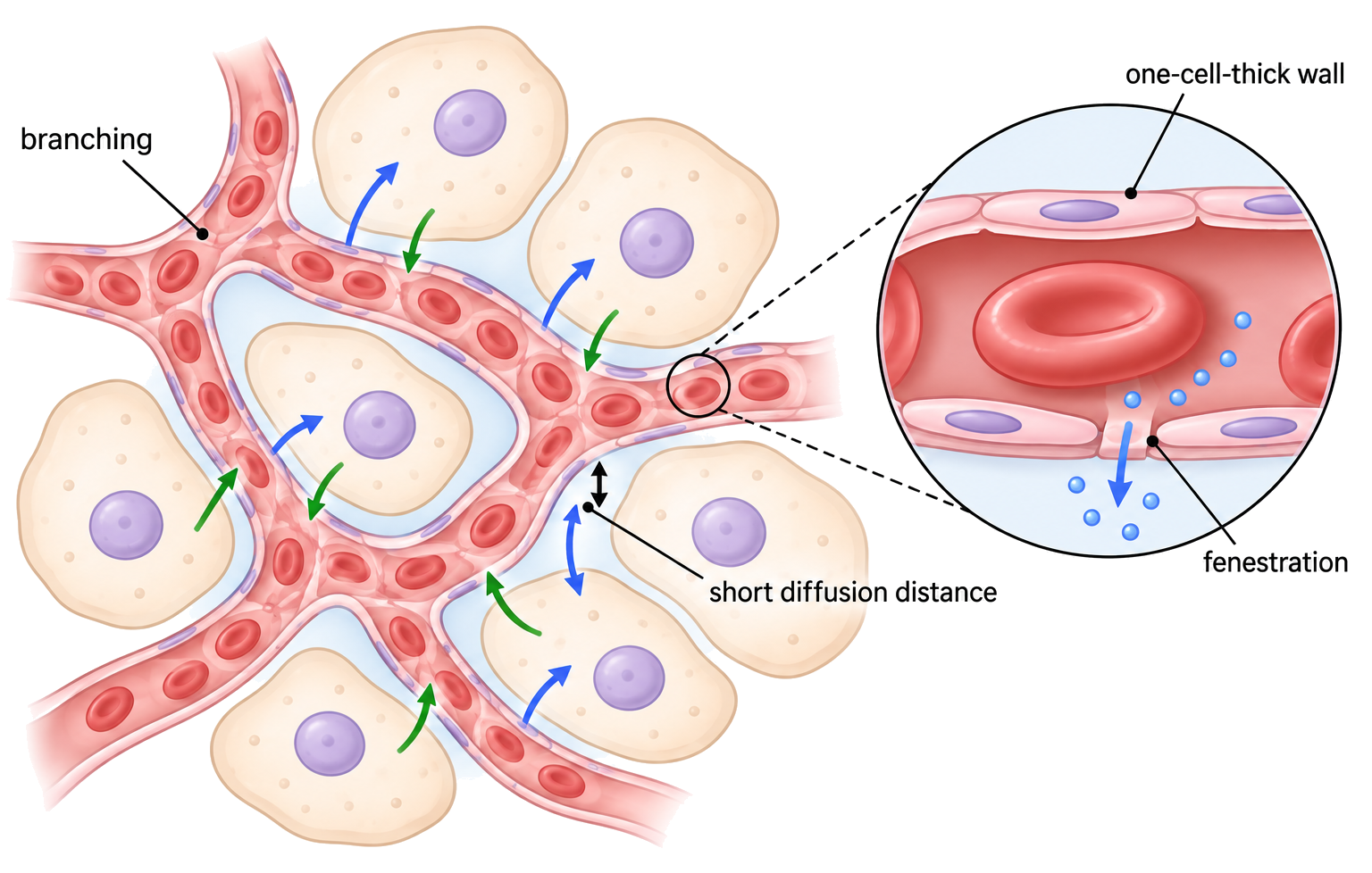
Branching produces a very large total surface area. A lumen only slightly wider than a red blood cell brings blood close to the wall, and a one-cell-thick endothelium provides a short path for diffusion.

Some capillaries have fenestrations—small pores through endothelial cells—where especially rapid fluid or solute exchange is required. Their narrow diameter also slows individual red cells and increases exchange time.

In an alveolar capillary, oxygen crosses thin alveolar and capillary layers into a red blood cell; in a fenestrated capillary, pores permit faster movement of water and small dissolved substances.

Thin walls suit exchange, not high-pressure transport. Fenestrations occur in some capillary beds, not every capillary, and blood cells plus most large proteins normally remain inside.

Capillary adaptations

Assessment in practice

1–2 marks
How it is assessed

This objective is assessed through structured response, commonly using Identify / Describe / Explain.

Command terms

Identify / Describe / Explain

What earns marks

Build the answer around this relationship: Capillary walls are one cell thick, giving a short diffusion path.

Watch for

Describing capillary walls as thin membranes instead of one-cell-thick endothelial walls.

Representative question

Question 1

[Maximum number: 3]

Explain how the structure of capillaries relates to their functions.

Arteries and Veins Handle Different Pressures

Arteries carry blood away from the heart under higher pressure; veins return blood at lower pressure and need valves and muscle assistance.

Thick elastic and muscular artery walls absorb pulse pressure. Veins have wider lumens, thinner walls and valves that prevent backflow as surrounding muscles compress them.

Trace direction and pressure first; then use wall thickness, lumen and valves to explain the vessel’s job.

During walking, leg muscles squeeze veins and push blood past valves toward the heart; the artery on the same route carries blood away under pulse pressure.

‘Away from the heart’ defines an artery, not oxygen content; the pulmonary artery carries deoxygenated blood.

Artery and vein structure

Assessment in practice

1–3 marks
How it is assessed

This objective is assessed through structured response, commonly using Identify / Label / Describe.

Command terms

Identify / Label / Describe / Explain / Distinguish

What earns marks

Build the answer around this relationship: Arteries have thicker walls and smaller lumens than veins.

Watch for

Using non-visible features such as valves when a micrograph question asks for visible artery-vein differences.

Representative question

Question 1

[Maximum number: 8]

Explain the structures and functions of arteries and veins.

Artery Walls Withstand Pulse Pressure

Arteries have thick muscular and elastic walls that maintain a lumen and smooth pressure pulses from the heart.

Elastic recoil helps keep blood moving between heartbeats, while smooth muscle adjusts diameter and resistance. The wall must withstand higher pressure than a vein.

Separate pulse smoothing from resistance control: which wall feature does each job?

A constricted arteriole raises resistance and can redirect blood flow, whereas elastic recoil in a large artery smooths the pulse.

A thick wall does not mean blood always flows faster; diameter, resistance and downstream demand also matter.

Artery adaptations

Assessment in practice

1–3 marks
How it is assessed

This objective is assessed through structured response, commonly using Identify / Explain.

Command terms

Identify / Explain

What earns marks

Build the answer around this relationship: Thick collagen-rich artery walls resist rupture under high pressure.

Watch for

Saying arteries pump blood by themselves rather than explaining elastic recoil after ventricular contraction.

Representative question

Question 1

[Maximum number: 5]

Explain how the structure of an artery allows it to carry out its function efficiently.

Pulse Rate Is a Repeated Pressure Signal

Pulse rate is the number of arterial pressure waves per minute, normally corresponding to ventricular contractions.

Place fingertips—not the thumb—lightly over the radial artery at the wrist or the carotid artery in the neck. Count waves for a known interval while the subject is still and use a full minute when maximum accuracy is needed.

Pulserate(beatsmin1)=pulsecount×(60s÷countingintervalins).Pulse rate (beats min⁻¹) = pulse count × (60 s ÷ counting interval in s).

Counting 18 pulses in 15 s gives 18 × (60/15) = 72 beats min⁻¹. Repeat after recovery and compare with a digital heart-rate sensor recorded over the same interval.

Short counts magnify counting error, and pulse rate is not cardiac output: cardiac output also depends on stroke volume. Record posture, activity and method when comparing results.

Pulse rate measurement

Assessment in practice

2 marks
How it is assessed

This objective is assessed through structured response, commonly using Outline.

Command terms

Outline

What earns marks

Build the answer around this relationship: Pulse is an arterial pressure wave produced by ventricular contraction.

Representative question

Question 1

[Maximum number: 2]

Outline one method that the researchers could have used to measure heart rate in this study.

Veins Return Blood at Low Pressure

Veins use large lumens, valves and skeletal-muscle contractions to return blood to the heart despite low pressure.
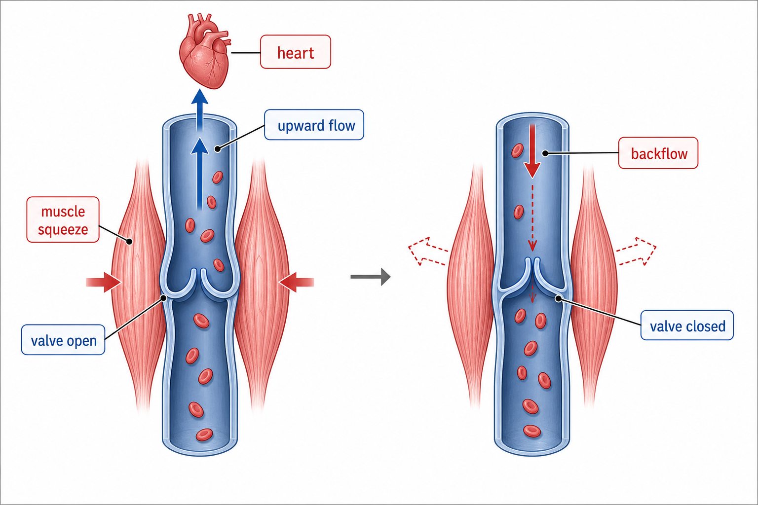
A wide lumen reduces resistance, valves stop reverse flow and muscle compression raises local pressure. Breathing movements can also help draw venous blood toward the chest.

Follow one bolus of venous blood upward and identify how valves and muscle compression prevent reversal.

When calf muscles contract, a valve below the compressed region closes while the valve above opens, pushing blood upward.

Valves do not create the original pressure; they make one-way assistance effective.

Vein adaptations

Assessment in practice

1–2 marks
How it is assessed

This objective is assessed through structured response, commonly using Identify / State / Explain.

Command terms

Identify / State / Explain / Deduce

What earns marks

Build the answer around this relationship: Vein valves keep blood moving toward the heart.

Watch for

Explaining venous flow without mentioning valves that prevent backflow.

Representative question

Question 1

[Maximum number: 2]

Deduce what the experiment demonstrated about the circulation of blood.

Coronary Occlusion Starves Heart Muscle

Coronary artery occlusion interrupts oxygen delivery to cardiac muscle and can cause myocardial infarction: irreversible death of part of the heart muscle.

Endothelial damage can allow lipid-rich atheroma to develop beneath the artery lining. A plaque narrows the lumen, and rupture can activate platelets and fibrin formation, producing a thrombus that partly or completely blocks flow.

Reduced coronary flow causes ischaemia: aerobic ATP production falls while cardiac muscle continues to demand energy. Prolonged complete occlusion damages and kills the supplied tissue, impairing contraction.

Epidemiological data may show a positive correlation between a proposed risk factor and coronary disease. A correlation coefficient quantifies direction and strength, but confounding variables and study design must be considered.

Even a strong correlation does not by itself prove that one variable causes coronary occlusion. Distinguish gradual plaque narrowing from an acute thrombus after plaque rupture.

Coronary artery occlusion

Assessment in practice

1–2 marks
How it is assessed

This objective is assessed through structured response, commonly using Outline.

Command terms

Outline

What earns marks

Build the answer around this relationship: Coronary arteries supply oxygen to cardiac muscle.

Watch for

Treating cholesterol correlation data as proof of direct causation.

Representative question

Question 1

[Maximum number: 2]

Outline the causes and consequences of blood clot formation in coronary arteries.

Roots and Xylem Form a Continuous Water Path

Transpiration creates tension—a negative pressure potential—that pulls a continuous water column from roots to leaves through xylem.

Water evaporating from moist mesophyll cell walls draws replacement water through the wall by capillary action and out of nearby xylem. This lowers pressure in the leaf xylem and transmits tension down the vessel.

Cohesion from hydrogen bonding keeps water molecules joined so the pull is transmitted through an unbroken column. Adhesion to hydrophilic xylem walls assists capillary movement and helps stabilize the column.

When stomata open and evaporation increases, the leaf water potential becomes more negative, increasing tension in xylem and drawing water upward from roots.

The main long-distance force is tension generated at transpiring leaves, not an active pump in xylem. Cohesion transmits the pull; it does not create the initial gradient.

Water transport in plants

Assessment in practice

2–7 marks
How it is assessed

This objective is assessed through structured response, commonly using Identify / Describe / Explain.

Command terms

Identify / Describe / Explain / Predict

What earns marks

Build the answer around this relationship: Transpiration pull creates tension that draws water upward through xylem.

Watch for

Explaining water movement as active transport through xylem rather than passive tension-driven flow.

Representative question

Question 1

[Maximum number: 8]

Explain the process of water uptake and transport by plants.

Xylem Tubes Carry Water Efficiently

Mature xylem vessel elements form dead, hollow, lignified tubes adapted to carry water under tension with little resistance.
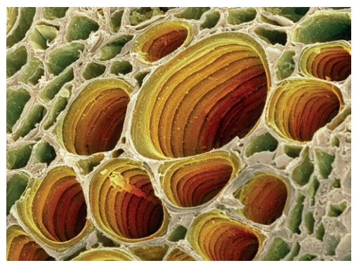
Loss of cytoplasm and organelles leaves an open lumen. End walls are absent or perforated, so aligned elements form a continuous route rather than forcing water across repeated membranes.

Lignin thickens and waterproofs the wall and prevents collapse under negative pressure. Unlignified pits allow water to enter or leave laterally, bypass a blockage, and move between xylem and surrounding tissue.

If air blocks one vessel, water can pass through pits into an adjacent vessel while lignified walls keep both tubes open under transpiration tension.

Xylem vessel elements are dead at maturity and do not actively pump water. Pits are thin wall regions for lateral movement, not open ends of the vessel.

Xylem vessel adaptations

Assessment in practice

2–3 marks
How it is assessed

This objective is assessed through structured response, commonly using Identify / Outline.

Command terms

Identify / Outline

What earns marks

Build the answer around this relationship: Xylem vessels lack cell contents, so water flow is less obstructed.

Watch for

Saying xylem is dead without explaining how lack of contents reduces resistance.

Representative question

Question 1

[Maximum number: 3]

Joints are part of the musculoskeletal system of animals, which provides support and movement to the body. Xylem provides support in plants and also transports water and minerals. Explain the adaptations of xylem for its functions.

Stem Tissues Place Transport in Separate Paths

A transverse section of a young dicot stem has an outer epidermis, cortex beneath it, vascular bundles arranged in a ring and a central pith.

Within each vascular bundle, phloem lies toward the outside, xylem toward the centre and cambium between them. The ring links transport around the stem while lignified xylem and supporting fibres add strength.

For a plan diagram, draw tissue boundaries with clear single lines and correct relative positions; do not draw individual cells. Label epidermis, cortex, vascular bundles, phloem, cambium, xylem and pith, then annotate their main functions.

Annotate xylem as water/mineral transport and support, phloem as translocation of assimilates, cortex as storage/support and epidermis as the protective outer boundary.

A plan diagram shows distribution and proportion, not cellular detail or shading. In each dicot stem bundle, xylem is inner and phloem outer; do not reverse them.

Stem tissue distribution

Assessment in practice

1 marks
How it is assessed

This objective is assessed through structured response, commonly using Identify / Label / Describe.

Command terms

Identify / Label / Describe

What earns marks

Build the answer around this relationship: Dicot stem vascular bundles are arranged in a ring.

Watch for

Confusing xylem and phloem positions within a dicot stem vascular bundle.

Representative question

Question 1

[Maximum number: 2]

Describe the distribution of vascular tissues in the stem of dicotyledonous plants.

Roots Combine Absorption with Selective Entry

A transverse section of a young dicot root has an outer epidermis, a broad cortex and a central vascular cylinder containing xylem and phloem.

Root hairs extend from epidermal cells to increase absorption area. The endodermis forms the inner boundary of the cortex, surrounding the central vascular tissue and controlling entry to xylem.

In the centre, xylem commonly forms a star or cross; phloem occurs in groups between its arms. Draw these tissue regions in their correct relative positions with clear outlines rather than individual cells.

A suitable plan diagram runs epidermis/root hairs → cortex → endodermis → central xylem cross with phloem between the arms, with annotations for uptake and transport.

Root vascular tissue is central rather than arranged as a ring of separate bundles like a young dicot stem. A plan diagram records tissue distribution, not detailed cell anatomy.

Root tissue distribution

Assessment in practice

1–2 marks
How it is assessed

This objective is assessed through structured response, commonly using Label.

Command terms

Label

What earns marks

Build the answer around this relationship: Dicot roots have central vascular tissue rather than a ring of separate bundles.

Watch for

Confusing the central root xylem arrangement with the ring of vascular bundles in stems.

Representative question

Question 1

[Maximum number: 2]

Label tissues X and Y .

Link Transport Structure To Function

Animal and plant transport answers should link structure to function. In animals, capillaries exchange, arteries maintain high-pressure flow, veins return low-pressure blood, pulse measures arterial pressure waves, and coronary occlusion blocks oxygen delivery to heart muscle. In plants, xylem transports water by transpiration tension and cohesion, while stem and root tissue plans show where xylem and phloem are arranged.

  • Blood vessel answers need structure plus pressure or exchange function.
  • Xylem answers need transpiration pull, cohesion, adhesion, lignin, pits, and hollow vessels when relevant.
  • Plant diagrams should identify tissue distribution: stem vascular bundles in a ring, root xylem cross with phloem between arms.

Objective notes

10 learning objectives
B3.2.1Capillary adaptations• Capillaries are narrow, highly branched, and close to cells• One-cell-thick endothelial walls reduce diffusion distance• Fenestrations in some capillaries allow rapid exchange and tissue fluid formation5% of analysed papers 7 papers · 7 questionsViewB3.2.2Artery and vein structure• Arteries and veins have endothelium, smooth muscle, elastic tissue, and collagen• Arteries have thicker walls and smaller lumens to withstand high pressure• Veins have wider lumens and thinner walls for low-pressure return6% of analysed papers 8 papers · 11 questionsViewB3.2.3Artery adaptations• Thick artery walls and collagen prevent rupture under high pressure• Elastic fibres stretch and recoil to even out pulse pressure and maintain flow• Smooth muscle in arteries and arterioles regulates blood distribution6% of analysed papers 8 papers · 8 questionsViewB3.2.4Pulse rate measurement• Ventricular contraction creates a pressure wave felt as a pulse• Pulse rate can be measured at radial or carotid arteries• Counting for a full minute is most accurate; shorter counts can be scaled0% of analysed papers ViewB3.2.5Vein adaptations• Veins have valves that prevent backflow toward capillaries• Thin flexible walls allow surrounding muscles to compress veins• Large lumens reduce friction during low-pressure blood return4% of analysed papers 5 papers · 5 questionsViewB3.2.6Coronary artery occlusion• Atherosclerosis forms plaques beneath damaged coronary artery endothelium• Plaque rupture can trigger thrombosis and occlude coronary arteries• Reduced oxygen supply can kill cardiac muscle and cause myocardial infarction1% of analysed papers 2 papers · 2 questionsViewB3.2.7Water transport in plants• Transpiration from leaf mesophyll creates tension in xylem water columns• Cohesion between water molecules transmits tension from leaves to roots• Adhesion to xylem walls helps maintain an unbroken transpiration stream1% of analysed papers 2 papers · 2 questionsViewB3.2.8Xylem vessel adaptations• Mature xylem vessels are dead, hollow tubes with absent or perforated end walls• Lignified walls resist collapse under negative pressure and waterproof the vessel• Pits allow lateral movement of water between vessels and surrounding tissues1% of analysed papers 1 paper · 1 questionViewB3.2.9Stem tissue distribution• Dicot stems have epidermis, cortex, pith, and vascular bundles in a ring• Each vascular bundle contains xylem, phloem, cambium, and supporting fibres• Plan diagrams show tissue positions without drawing individual cells0% of analysed papers ViewB3.2.10Root tissue distribution• Dicot roots have epidermis with root hairs, cortex, endodermis, and central vascular tissue• Xylem forms a central cross with phloem between its arms• The Casparian strip blocks apoplast flow and forces selective symplast entry to xylem1% of analysed papers 1 paper · 1 questionView