EduNinja

IGCSE Biology Extended14.2.2—Structures of the eyeTopic Practice

14.2.2—Structures of the eye

• Identify in diagrams and images the structures of the eye, limited to: cornea, iris, pupil, lens, retina, optic nerve and blind spot

Question 1(b)(i)

[Maximum number: 1]

Accommodation (focusing) is one of the functions of the eye.

Fig. 1.1 is a diagram of an eye that is focusing on a distant object.
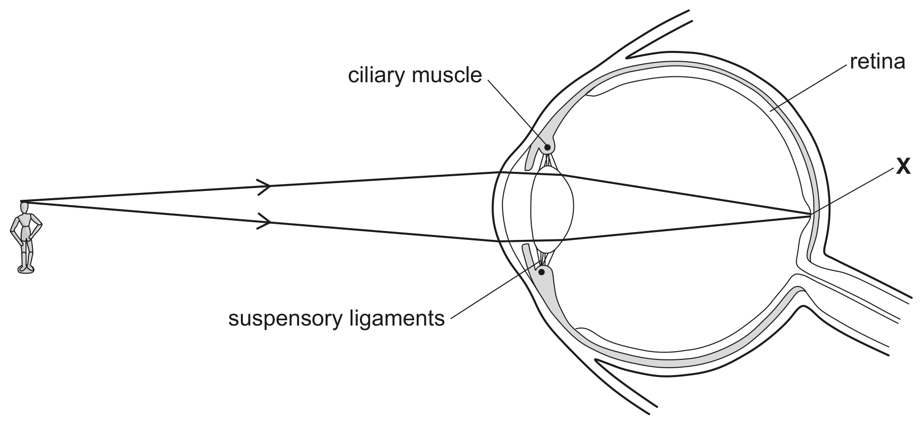
Fig. 1.1

Fig. 1.1

State the name of the part of the retina labelled X.

Fig. 1.2 is an incomplete diagram of an eye that is focusing on a near object.
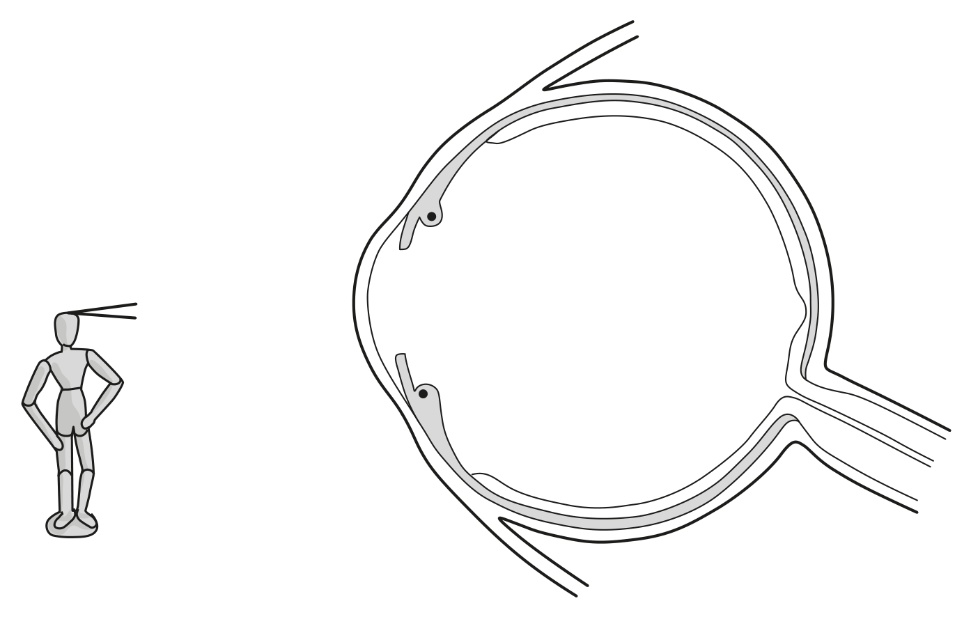
Fig. 1.2
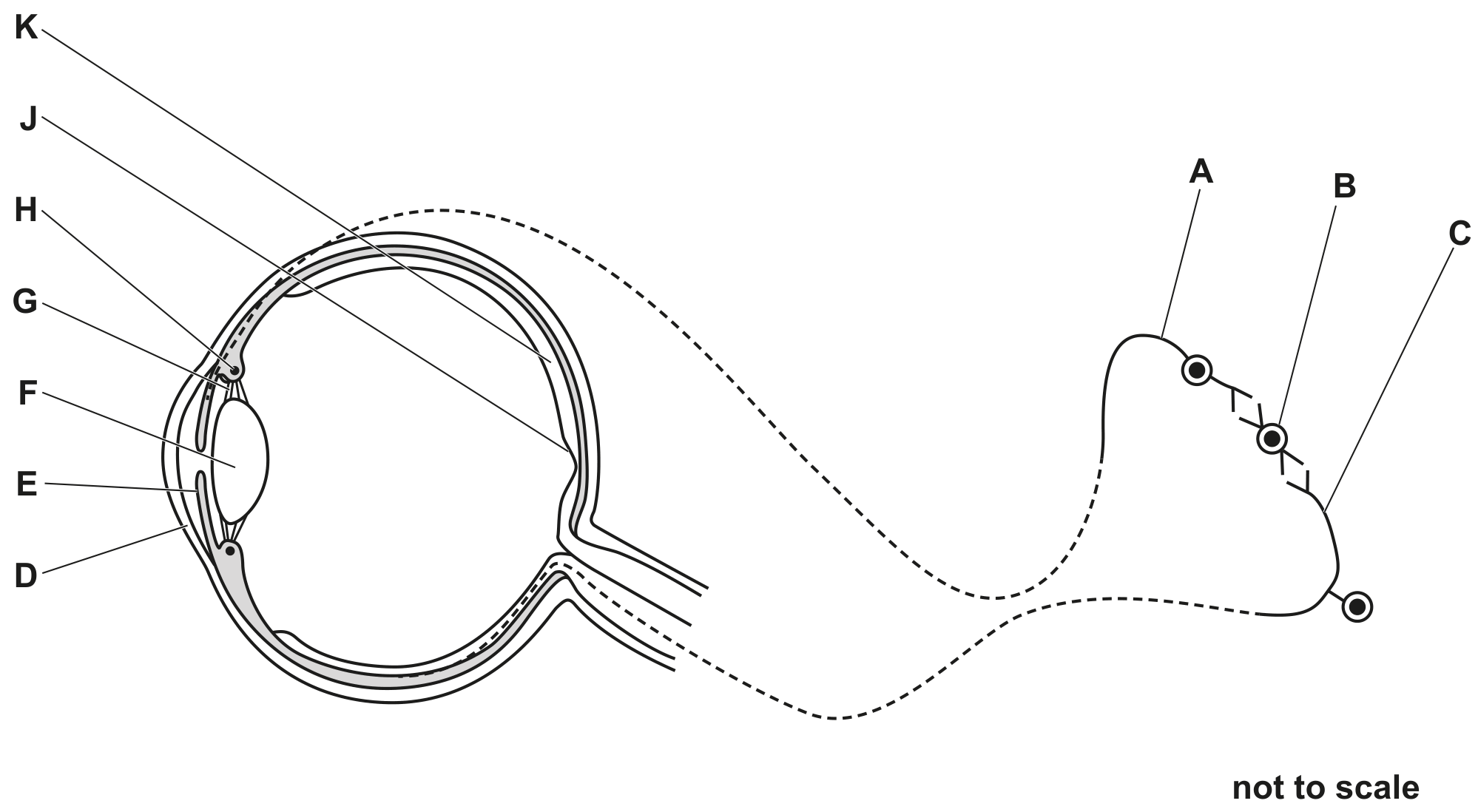
Fig. 1.2

Question 1(b)(ii)

[Maximum number: 4]

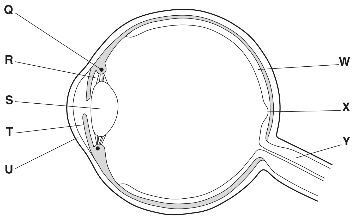
Fig. 1.1 shows part of a human eye and three neurones that conduct electrical impulses between the eye and the brain. These neurones are involved in the pupil reflex.

Fig. 1.1

Fig. 1.1

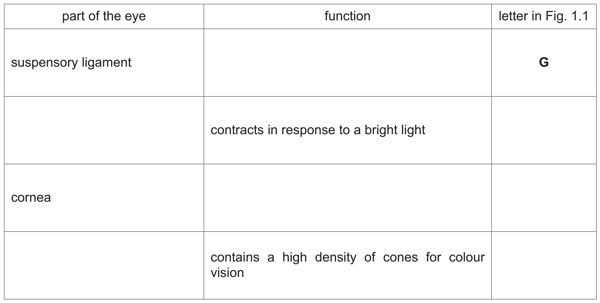
Table 1.1 shows the names of some parts of the eye, their functions and the letters in Fig. 1.1 that identify the parts of the eye.

Complete Table 1.1.

Table 1.1

Table 1.1

Question 2

[Maximum number: 2]

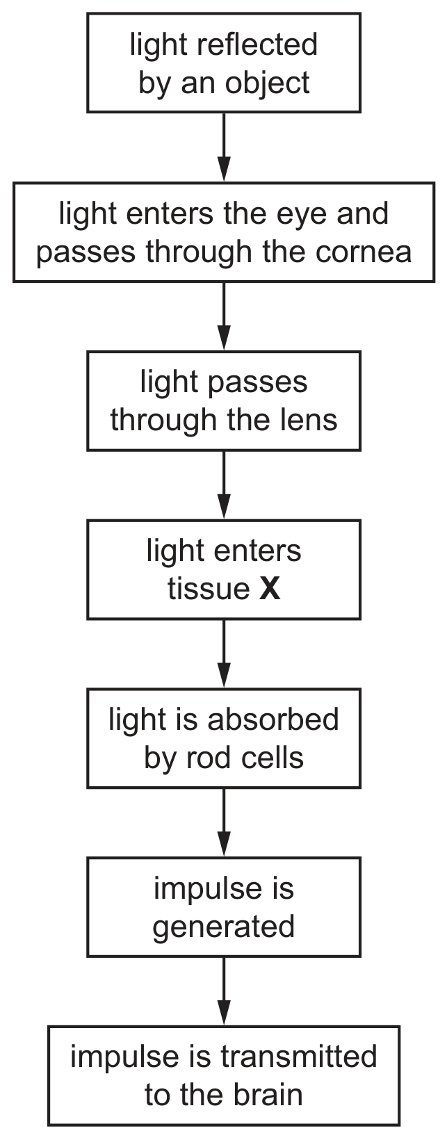
Fig. 2.1 is a flow chart that shows the events that occur as light travels through the eye.

Fig. 2.1

Fig. 2.1

Question 2(a)(i)

(a)

State the name of the tissue X.

[ 1 ]

Question 2(a)(ii)

(b)

State the name of the nerve that transmits impulses from the eye to the brain.

[ 1 ]

Question 3

Question 3(a)(ii)

(a)

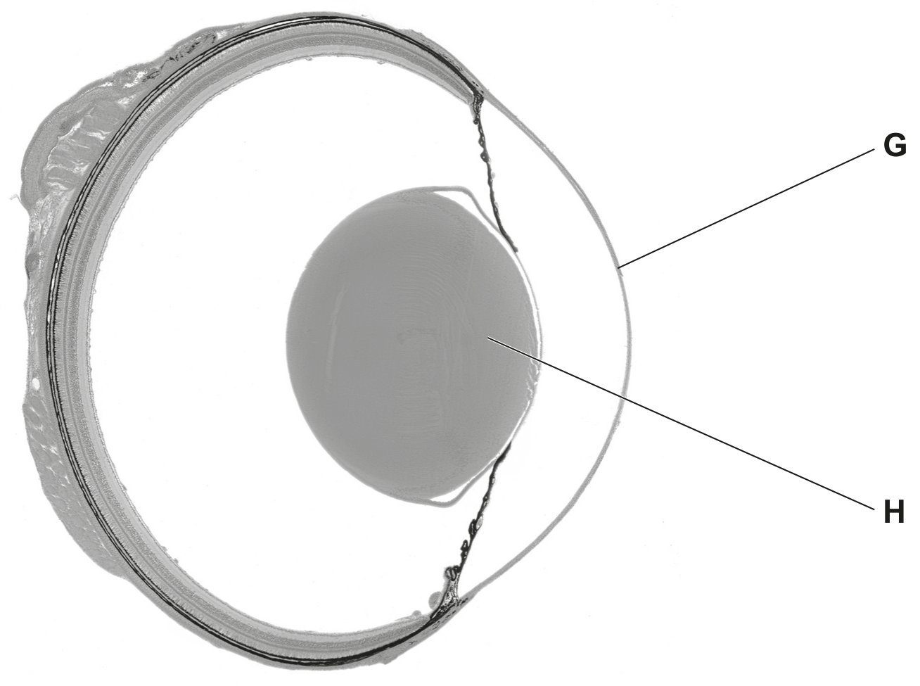
Fig. 3.1 is a photograph of a cross-section of a frog's eye.

Fig. 3.1

Fig. 3.1

Identify the structures labelled G and H in Fig. 3.1.
G

H

[ 2 ]

Question 3(c)

(b)

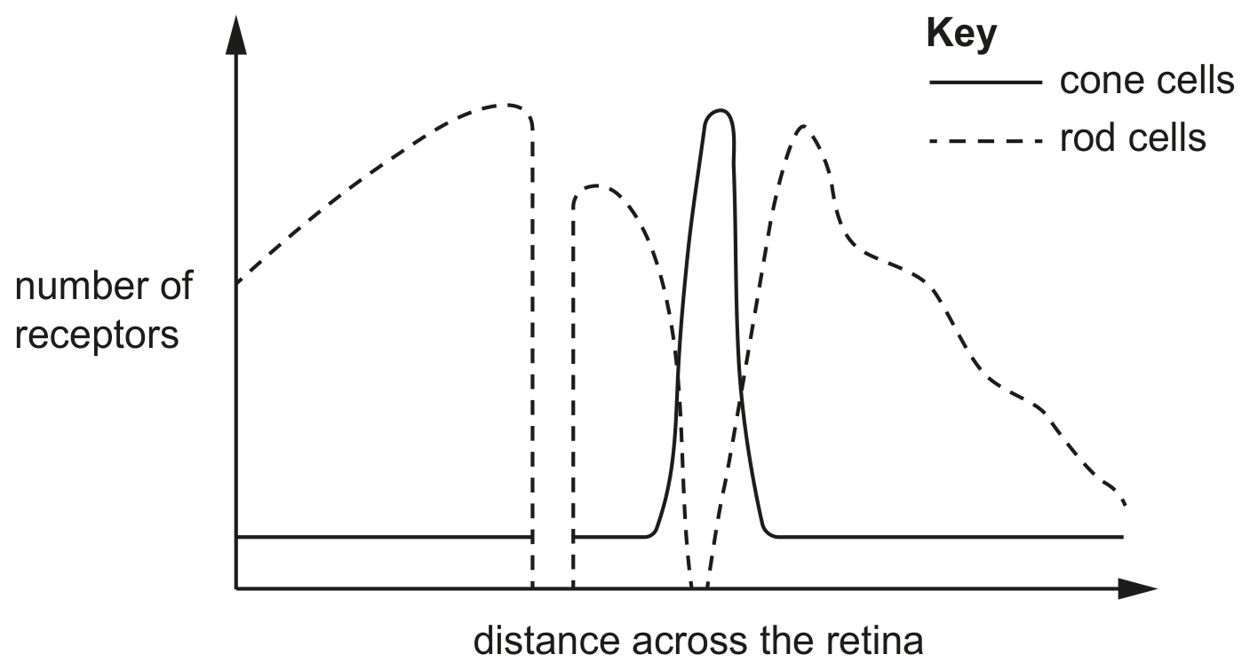
Fig. 3.3 is a graph showing the distribution of rod cells and cone cells across the retina in a human eye.

Fig. 3.3

Fig. 3.3

[ 2 ]

Question 3(c)(i)

(i)

On Fig. 3.3 draw:
- the letter P to show the location of the fovea
- the letter Q to show the location of the blind spot.

[ 2 ]

Question 4

Question 4(b)

(a)

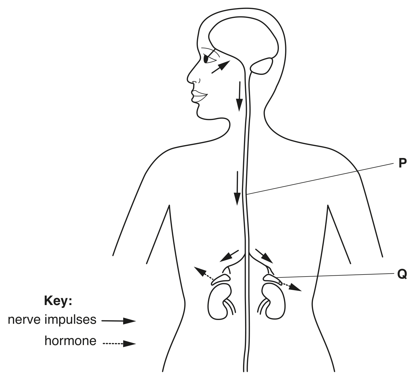
The responses of the human body to danger are coordinated by the nervous and endocrine systems.

Fig. 4.1 shows the sequence of events that occurs in response to a dangerous situation that is detected by the eyes.

Fig. 4.1

Fig. 4.1

[ 2 ]

Question 4(b)(iv)

(i)

State the nerve that contains these neurones that conduct impulses from the eye to the brain.

[ 1 ]

Question 4(b)(v)

(ii)

Identify the organ labelled P.

[ 1 ]

Question 24

[Maximum number: 1]

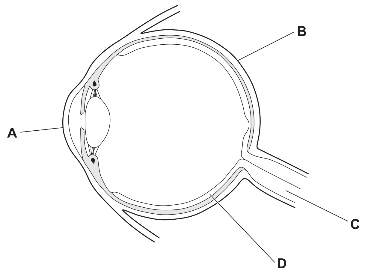
The diagram shows an eye.
Which labelled part contains cells that are sensitive to light?

Question image

Question 4(a)

[Maximum number: 5]

The eye is a sense organ that responds to light.
Fig. 4.1 is a diagram of a section through the human eye.

Fig. 4.1

Fig. 4.1

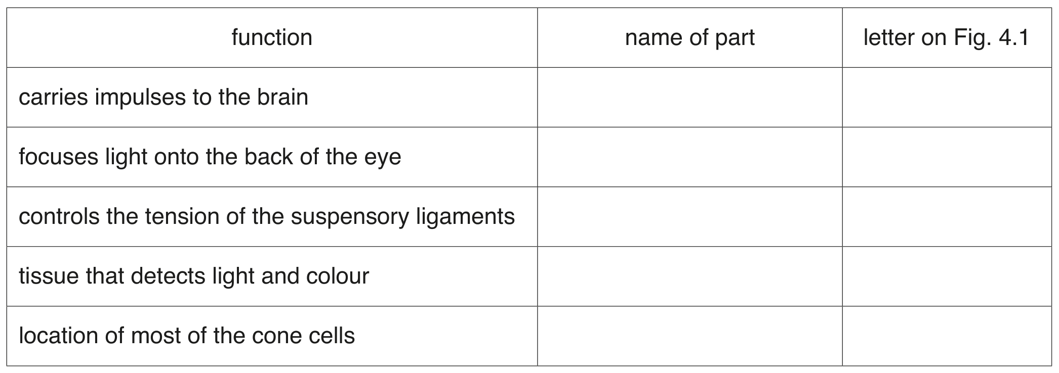
Table 4.1 describes some of the functions of the parts of the eye.

Complete the table by:
- naming the parts of the eye
- using the letters on Fig. 4.1 to identify the parts of the eye.

Table 4.1

Table 4.1

Question 26

[Maximum number: 1]

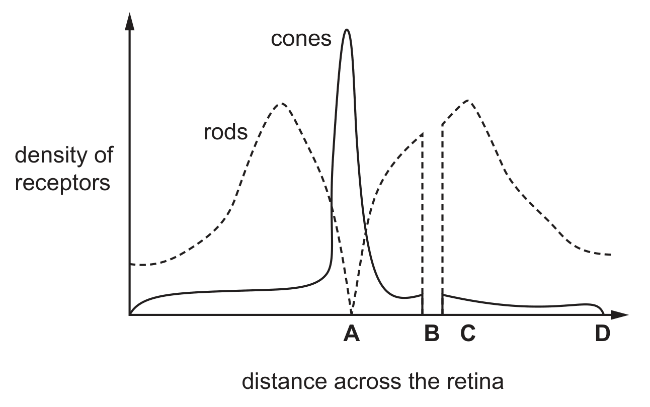
The diagram shows the density of rods and cones across a horizontal section of the retina.
What is the position of the optic nerve?

Question image

Question 26

[Maximum number: 1]

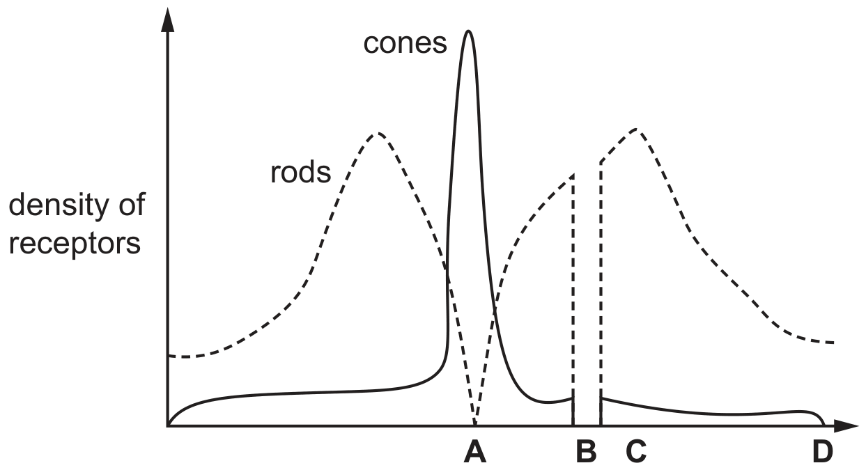
The diagram shows the density of rods and cones across a horizontal section of the retina.
What is the position of the optic nerve?

Question image

distance across the retina

Question 4(a)

[Maximum number: 1]

Hydrophytes are plants that show many adaptive features for life in aquatic habitats.
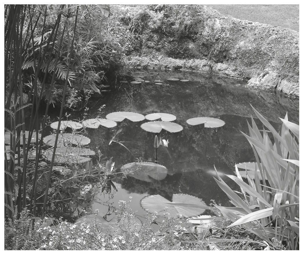
Fig. 4.1 shows several species of hydrophyte growing in freshwater.

Fig. 4.1

Fig. 4.1

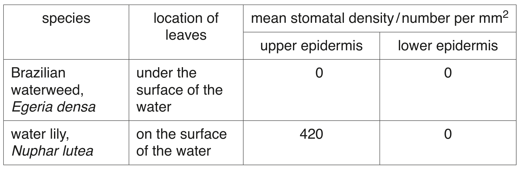
A student investigated the density of stomata on the leaves of two different species of freshwater hydrophyte.

Table 4.1 shows the results.

Table 4.1

Table 4.1

Name the epidermal cells that control the size of stomata.

0 selected