A-Level Biology Cell Structure: AS Revision Guide
Revise A-Level Biology cell structure with organelles, prokaryotic vs eukaryotic cells, microscopy, magnification, and cell adaptation wording.
If A-Level Biology cell structure feels like a long organelle list, start with a different question: what would the examiner expect you to recognise, compare, measure, or explain?
In AS Biology, this topic usually tests two things together. You need the microscope skills, especially magnification, resolution, and units. You also need the cell content: plant and animal cells, eukaryotic organelles, prokaryotic cells, and the basic structure of viruses.
This guide follows the EduNinja source PDF, AS Biology Revision Notes - Cell Structure. Use it as a revision map rather than a separate set of facts to memorise.
Useful starting points:
- A-Level Biology Notes
- A-Level Biology Question Bank
- AS Biology Revision Notes - Cell Structure
- AS Biology Revision Notes - Cell membranes and transport
Quick answer
For A-Level Biology cell structure, revise these first:
- Magnification means how much bigger the image appears than the real specimen.
- Resolution means the ability to distinguish two separate points as separate.
- Convert units before using a magnification formula: 1 mm = 1000 um and 1 um = 1000 nm.
- Light microscopes have lower resolution than electron microscopes, but they can be used with living tissue.
- Plant cells have a cellulose cell wall, plasmodesmata, and a large permanent vacuole. Many plant cells, especially photosynthetic cells, contain chloroplasts.
- Animal cells usually have centrioles near the nucleus, inside a region called the centrosome.
- Eukaryotic cells have a nucleus and membrane-bound organelles.
- Prokaryotic cells lack a nucleus and membrane-bound organelles, but they still have 70S ribosomes and circular DNA.
- Viruses are acellular. They have a protein coat and a nucleic acid core, and they replicate only inside host cells.
If you only remember one thing, do not revise organelles as isolated labels. Pair every structure with a precise function and the kind of evidence you might see in a diagram, micrograph, or comparison question.
What cell structure includes in AS Biology
The source PDF splits cell structure into two connected areas:
| Area | What the PDF covers | What students should practise |
|---|---|---|
| The microscope in cell studies | plant and animal cell images, magnification, resolution, light microscopes, electron microscopes | calculations, unit conversion, microscope comparison, drawing and labelling |
| Cells as the basic units of living organisms | eukaryotic organelles, plant structures, prokaryotic cells, viruses | structure-function answers, comparison tables, diagram recognition |
The topic also prepares you for later chapters. Mitochondria link to respiration. Chloroplasts link to photosynthesis. Ribosomes, rough ER, and the Golgi body link to protein synthesis and secretion. The cell surface membrane links to transport.
That is why a good answer does not stop at naming the organelle. It says what the structure does and, where needed, how that function supports the cell.
Microscope skills: magnification, resolution, and units
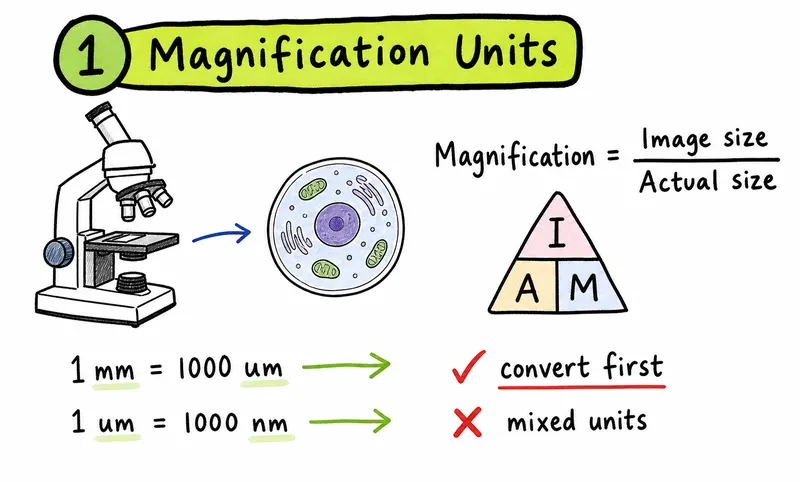
The PDF defines magnification as how much bigger a sample appears under a microscope than it is in real life. Resolution is different. It is the ability to distinguish two separate points.
Use these formulas:
| Formula | Use |
|---|---|
| magnification = image size / actual size | when comparing the drawn or photographed size with the real size |
| actual size = image size / magnification | when the question gives a magnified image |
| image size = actual size x magnification | when predicting the size of a drawn or displayed image |
Check units before calculating:
| Conversion | Meaning |
|---|---|
| 1 mm = 1000 um | useful for microscope drawings and cell measurements |
| 1 um = 1000 nm | useful for membranes, ribosomes, and electron micrographs |
A common mistake is to calculate with mixed units, such as using mm for image size and um for actual size. Convert first, then substitute.
Light and electron microscopes
The source PDF gives a clear comparison between light microscopes and electron microscopes:
| Microscope type | Approximate resolution | Approximate magnification |
|---|---|---|
| Light microscope | 200 nm | x1500 |
| SEM | 3 nm | high electron microscope magnification |
| TEM | 0.5 nm | x250,000 to x500,000 |
Electron microscopes have much higher resolving power, so they can show more internal cell detail. Ribosomes are about 25 nm, which is why they cannot be seen clearly with a light microscope.
Light microscopes still matter. According to the PDF, they have several advantages over electron microscopes:
| Light microscope advantage | Why it matters |
|---|---|
| Can observe living tissue | living processes can be watched |
| More portable | easier to use in a school or lab setting |
| Easier to use | less technical training is needed |
| Can show real or natural colours | electron micrographs do not show natural colour |
| Stains can improve visibility | different stains can highlight different cell structures |
Electron microscopes need a vacuum because electrons scatter if they collide with air molecules. Water boils at room temperature in a vacuum, so the sample must be dehydrated. That means the specimen is dead.
For exam questions, say both sides: electron microscopes give higher resolution and magnification, while light microscopes can be used with living specimens and are easier to handle.
Plant and animal cells: the comparison to know
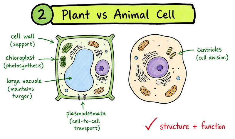
Plant and animal cells are both eukaryotic, so they share a nucleus, cytoplasm, a cell surface membrane, mitochondria, ribosomes, rough ER, smooth ER, and the Golgi body. Plant cells also contain structures that animal cells do not usually have.
| Structure | Plant cell | Animal cell | Exam wording to use |
|---|---|---|---|
| Cell wall | Present, made of cellulose | Absent | gives shape and support, freely permeable |
| Chloroplasts | Present in photosynthetic cells | Absent | site of photosynthesis, contains starch grains, circular DNA, and 70S ribosomes |
| Vacuole | Large permanent vacuole surrounded by tonoplast | Usually absent or smaller | contains cell sap and helps regulate osmotic properties |
| Plasmodesmata | Present between neighbouring plant cells | Absent | cytoplasmic connections for movement and communication |
| Centrioles | Usually absent in higher plant cells | Present in many animal cells | help organise microtubules and produce spindle fibres |
Do not write only "plant cells have a cell wall." A stronger comparison links the structure to function. For example, a cellulose cell wall gives support and helps prevent a plant cell from bursting when water enters.
Eukaryotic organelles and their functions
The PDF lists the main eukaryotic structures with their sizes and functions. Use the table below as a concise revision version.
| Structure | Source detail to remember | Function |
|---|---|---|
| Cell surface membrane | phospholipid bilayer, about 7 nm | selectively permeable barrier between cytoplasm and external environment; involved in cell recognition |
| Nucleus | about 7 um, surrounded by a nuclear envelope | controls cell activities and contains genetic material |
| Nuclear pores | openings in the nuclear envelope | control movement into and out of the nucleus, including mRNA leaving and proteins entering |
| Nucleolus | about 2.5 um | involved in ribosome synthesis |
| Ribosomes | about 25 nm, made of two subunits | site of protein synthesis |
| Rough ER | cisternae with ribosomes attached | proteins enter the sacs and may be modified as they pass through |
| Smooth ER | no attached ribosomes | synthesises lipids and steroids such as cholesterol and reproductive hormones |
| Golgi body | stack of flattened sacs formed from vesicles | modifies and packages substances into vesicles |
| Lysosomes | 0.1 to 1 um, single membrane | contain hydrolytic enzymes for breaking down unwanted structures |
| Mitochondria | 0.5 to 10 um | site of aerobic respiration and ATP synthesis |
| Microtubules | hollow tubes made from tubulin | support the cell and help maintain shape |
| Chloroplasts | 3 to 10 um | site of photosynthesis and ATP production in plant cells |
| Cell wall | cellulose, freely permeable | gives shape, support, and protection from bursting |
| Plasmodesmata | cytoplasmic strands through cell walls | allow movement of water, sucrose, amino acids, ions, and signals between plant cells |
| Vacuole | surrounded by a partially permeable tonoplast | contains cell sap and helps regulate the osmotic properties of the cell |
The PDF also notes that ribosomes in the cytoplasm are 80S, while ribosomes in chloroplasts and mitochondria are 70S. That detail is useful when you compare eukaryotic organelles with prokaryotic cells.
The protein pathway: ribosome to Golgi
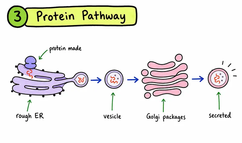
Several organelles in this topic fit into one sequence. This is one of the easiest ways to turn a list into an exam answer.
| Step | Structure | What happens |
|---|---|---|
| 1 | Ribosome on rough ER | protein synthesis occurs |
| 2 | Rough ER cisternae | proteins enter the sacs and may be modified |
| 3 | Vesicles | small sacs break off from the ER |
| 4 | Golgi body | proteins are modified, packaged, and sent out in vesicles |
The PDF gives examples of Golgi functions: glycosylation, phosphorylation of proteins, folding proteins, assembly of polypeptides into quaternary structure, and removing the first amino acid methionine to activate proteins.
You do not need to write all of those every time. Use them when the question asks for more detail about protein processing or secretion.
Mitochondria, chloroplasts, and ATP
Mitochondria carry out aerobic respiration. The PDF describes ATP as the energy-carrying molecule in all living cells. Energy from molecules such as sugars and fats is transferred to ATP during respiration. When ATP breaks down to ADP in a hydrolysis reaction, energy is released for cell processes.
Cells with high energy demand, such as muscle cells, liver cells, and root hair cells, often contain more mitochondria. The outer membrane of a mitochondrion contains a transport protein called porin.
Chloroplasts carry out photosynthesis. The PDF notes that chloroplasts contain starch grains, circular DNA, and 70S ribosomes. ATP is also produced in chloroplasts.
Use this distinction:
| Organelle | Main process | Good exam wording |
|---|---|---|
| Mitochondrion | aerobic respiration | synthesises ATP for cellular processes |
| Chloroplast | photosynthesis | contains chlorophyll-containing structures for photosynthesis and can produce ATP |
Avoid saying mitochondria "make energy." They transfer energy to ATP during aerobic respiration.
Prokaryotic cells: small but not empty
The PDF describes prokaryotes as organisms that lack nuclei or proper nuclear membranes. They are unicellular and usually about 1 to 5 um in size.
| Feature | Prokaryotic cell detail from the PDF |
|---|---|
| Nucleus | absent |
| Genetic material | circular DNA |
| Organelles | no membrane-bound organelles and no ER |
| Ribosomes | 70S ribosomes |
| Cell wall | made of murein, also called peptidoglycan |
| Size | about 1 to 5 um |
The exam trap is the phrase "prokaryotes have no organelles." That is too broad. Prokaryotes have ribosomes, but they do not have membrane-bound organelles such as mitochondria, chloroplasts, ER, or a Golgi body.
Use the comparison below for short-answer questions:
| Feature | Eukaryotic cell | Prokaryotic cell |
|---|---|---|
| Nucleus | present | absent |
| DNA | inside nucleus | circular DNA, not enclosed in a nucleus |
| Ribosomes | 80S in cytoplasm | 70S |
| Membrane-bound organelles | present | absent |
| Size | larger | smaller, about 1 to 5 um |
| Cell wall | cellulose in plant cells | murein or peptidoglycan in bacteria |
Viruses: what the PDF adds
The source PDF also includes viruses at the end of the topic. Viruses are not cells. They are acellular, with a protein coat and a nucleic acid core made from DNA or RNA.
| Virus feature | Detail |
|---|---|
| Cellular status | non-cellular or acellular |
| Outer structure | protein coat |
| Genetic material | DNA or RNA strand |
| Replication | only inside host cells |
| Living characteristics | show no characteristics of living organisms outside a host cell |
| Shape | often symmetrical |
The virus DNA or RNA takes over the protein-synthesising machinery of the host cell, which helps produce new virus particles.
This is a useful comparison point. Eukaryotic and prokaryotic cells both have cellular organisation. Viruses do not.
Common mistakes that cost marks
| Mistake | Better answer habit |
|---|---|
| Confusing magnification and resolution | Define magnification as image enlargement and resolution as distinguishing two separate points |
| Mixing mm, um, and nm | Convert all measurements before calculating |
| Saying ribosomes are visible with a light microscope | Remember ribosomes are about 25 nm and cannot be seen clearly with a light microscope |
| Saying electron microscopes can view living cells | Electron microscopes require a vacuum and dehydrated dead specimens |
| Saying mitochondria make energy | Say they are the site of aerobic respiration and ATP synthesis |
| Saying prokaryotes have no organelles | Say they lack membrane-bound organelles but have 70S ribosomes |
| Treating viruses as cells | Say viruses are acellular and replicate only inside host cells |
When you practise, turn each vague answer into one structure-function sentence. That is the fastest way to improve this topic.
A 30-minute revision route for cell structure
| Time | Task |
|---|---|
| 0-5 min | Learn the microscope definitions and unit conversions |
| 5-10 min | Redraw a plant cell and an animal cell from memory |
| 10-16 min | Build a structure-function table for 8 organelles |
| 16-21 min | Compare prokaryotic and eukaryotic cells |
| 21-26 min | Do one magnification calculation and one microscope comparison question |
| 26-30 min | Mark answers and rewrite every vague phrase |
If you want the loop in one place, use EduNinja Notes for the content, the Question Bank for exam-style practice, and flashcards for structure-function pairs you keep missing.
Question-type breakdown
| Question type | What it tests | First move |
|---|---|---|
| Magnification calculation | formula and unit conversion | convert units, then use image size, actual size, and magnification |
| Microscope comparison | resolution, magnification, living specimens, sample preparation | state one advantage and one limitation of each microscope |
| Organelle function | precise structure-function recall | name the organelle and give a specific function |
| Plant vs animal cell | visible differences and function | identify cell wall, chloroplasts, vacuole, plasmodesmata, and centrioles |
| Eukaryotic vs prokaryotic cell | cell organisation | compare nucleus, DNA, ribosomes, cell wall, and membrane-bound organelles |
| Virus comparison | acellular structure | mention protein coat, nucleic acid core, and replication inside host cells |
Weak answer vs mark-worthy answer
Weak:
- Mitochondria make energy.
Better:
- Mitochondria are the site of aerobic respiration, where energy is transferred to ATP.
Weak:
- Electron microscopes are stronger.
Better:
- Electron microscopes have higher resolution than light microscopes, so they can show smaller cell structures in more detail.
Weak:
- Prokaryotes have no organelles.
Better:
- Prokaryotes lack membrane-bound organelles, but they still contain 70S ribosomes.
Weak:
- Viruses are tiny cells.
Better:
- Viruses are acellular. They contain a protein coat and DNA or RNA, and they replicate only inside host cells.
Related Study Links
FAQ
What is the difference between magnification and resolution?
Magnification is how much larger the image appears than the real specimen. Resolution is the ability to distinguish two separate points. A microscope can produce a large image, but if the resolution is poor, the image will still lack clear detail.
Why can electron microscopes show more detail than light microscopes?
Electron microscopes have much higher resolution than light microscopes. The source PDF lists light microscope resolution at about 200 nm, SEM resolution at about 3 nm, and TEM resolution at about 0.5 nm. This allows electron microscopes to show smaller structures.
Why can light microscopes be useful if electron microscopes are more powerful?
Light microscopes can be used with living tissue, are more portable, and are easier to use. They can also show natural colours or stained specimens. Electron microscopes need a vacuum and dehydrated dead samples.
What is the difference between eukaryotic and prokaryotic cells?
Eukaryotic cells have a nucleus and membrane-bound organelles. Prokaryotic cells lack a nucleus and membrane-bound organelles, but they still have cell membranes, cytoplasm, circular DNA, and 70S ribosomes.
Are viruses cells?
Viruses are not cells. They are acellular particles with a protein coat and a DNA or RNA core. They replicate only inside host cells by using the host cell's protein-synthesising machinery.
Related study links
-
A-Level Biology Cell Structure: Organelles and Microscopy Notes
-
A-Level Biology Cell Structure: Organelles and Microscopy Question Bank
-
Revise from AS Biology Revision Notes - Cell Structure
-
Practise with the A-Level Biology Question Bank
-
Review A-Level Biology Notes
-
Connect the topic to transport with AS Biology Revision Notes - Cell membranes and transport
-
Study the next related article: A-Level Biology biological molecules and food tests
Put cell structure back into your full A-Level CAIE Biology AS study plan.
Return to the complete course overview, or move directly into the most relevant study tool.
Related articles
More course notes, updates and study resources from the EduNinja blog.
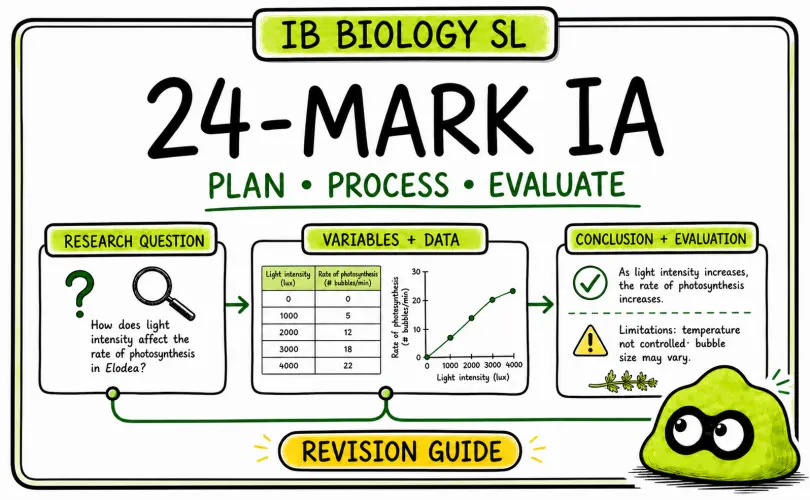
IB Biology Scientific Investigation Guide: How to Build a Strong 24-Mark IA
A practical IB Biology guide to the 24-mark Scientific Investigation: choose a workable research question, control variables, process data, draw a supported conclusion and evaluate limitations with specific improvements.
Abiotic Factors Explained | IB Biology SL
Revise IB Biology SL abiotic factors, tolerance ranges, limiting factors and how environmental variables affect distribution.
Active Transport vs Passive Transport | A-Level Biology
Revise A-Level Biology active and passive transport with concentration gradients, ATP, carrier proteins, examples, common mistakes and practice.