[Maximum number: 2]
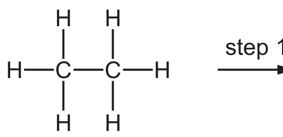
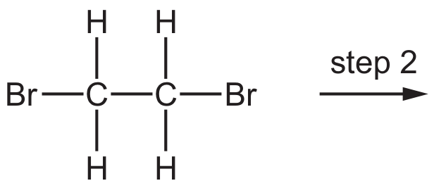
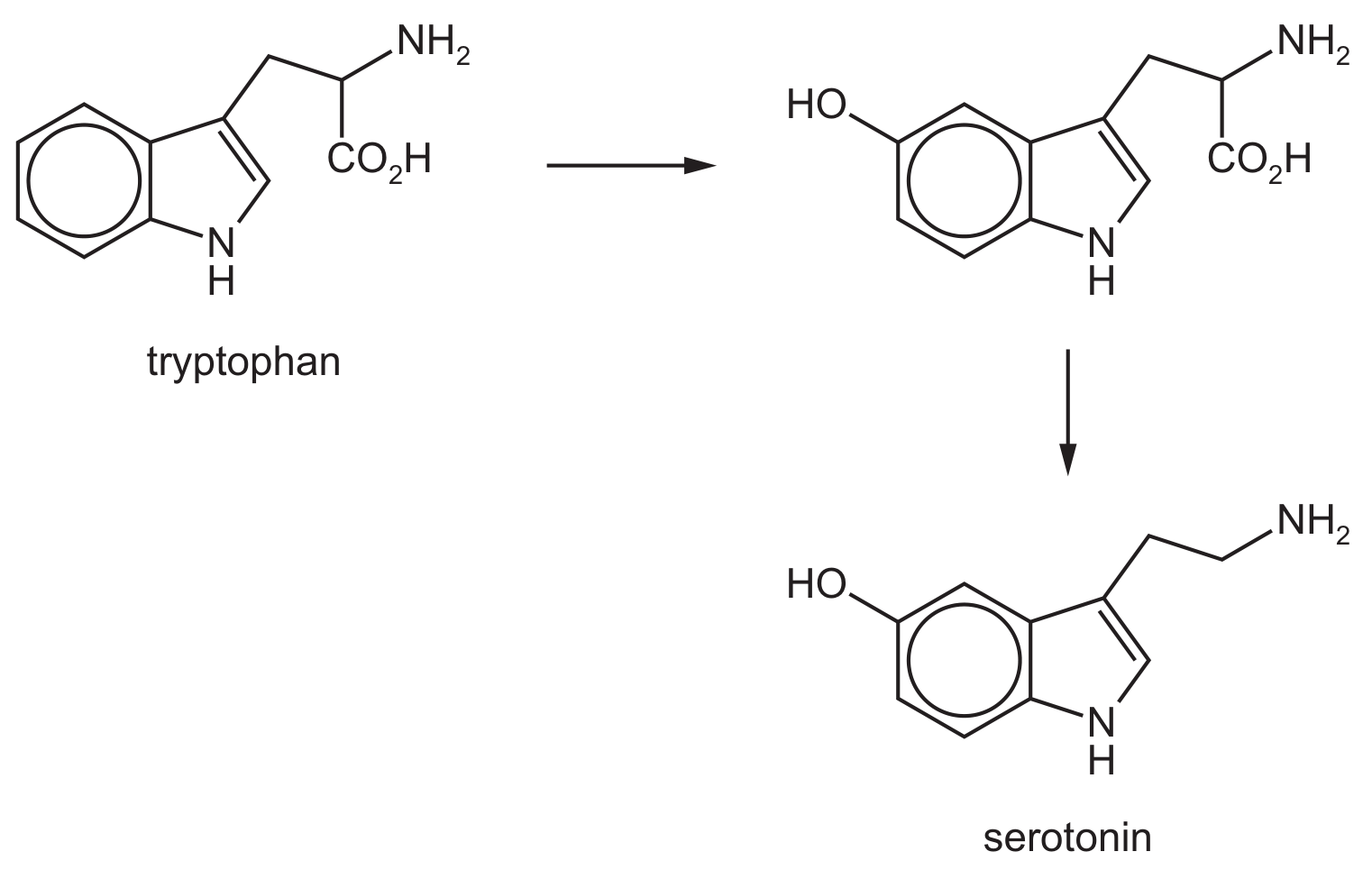
Serotonin can be synthesised from the amino acid tryptophan in two steps.
(a)
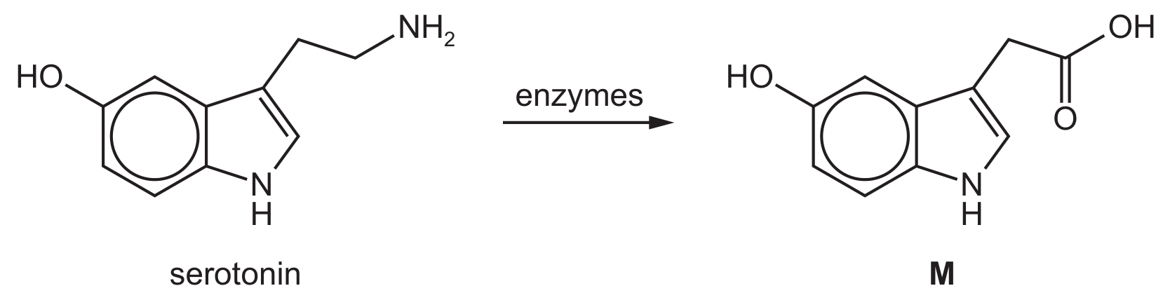
Serotonin is converted by enzymes in the liver to compound M.
[ 2 ]
(i)
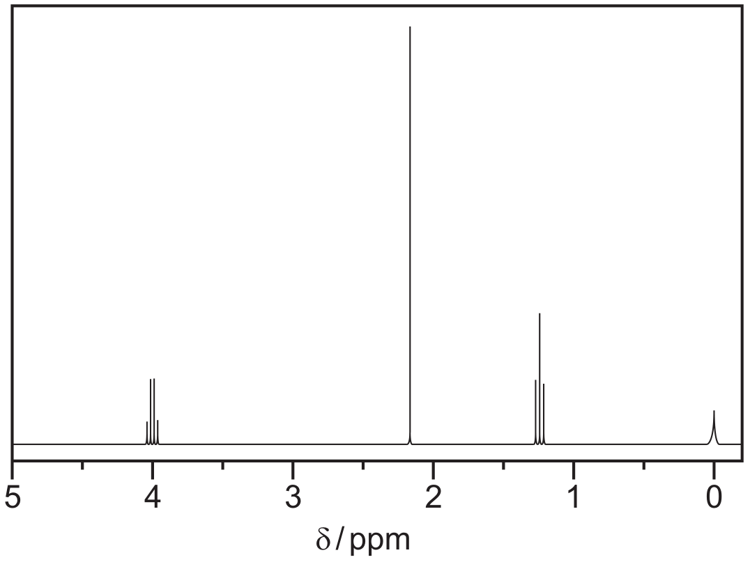
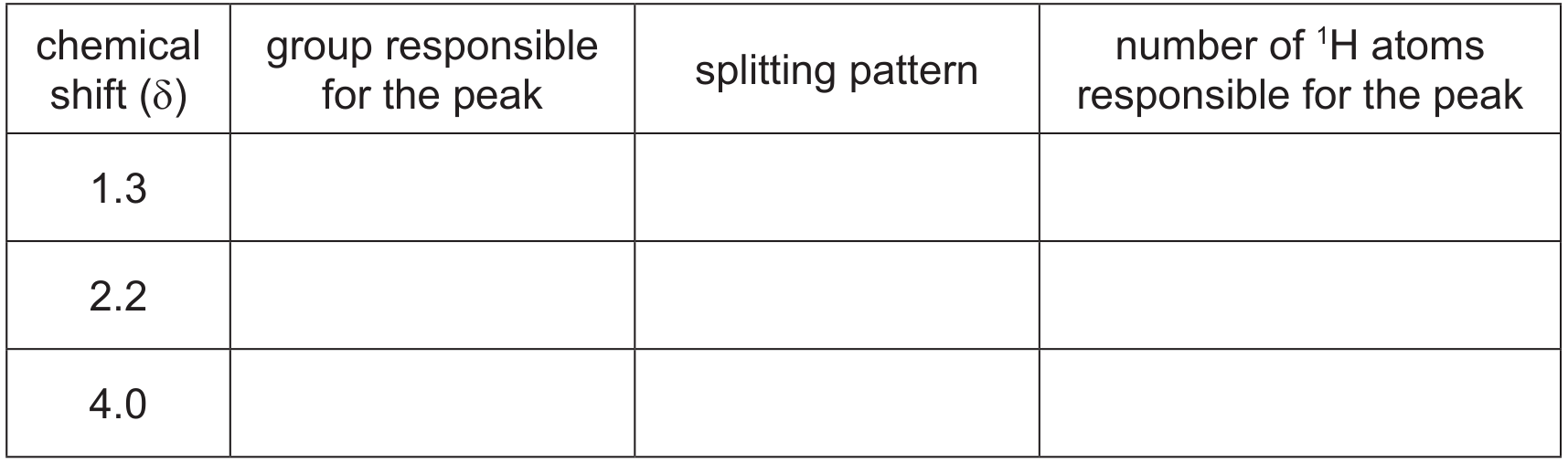
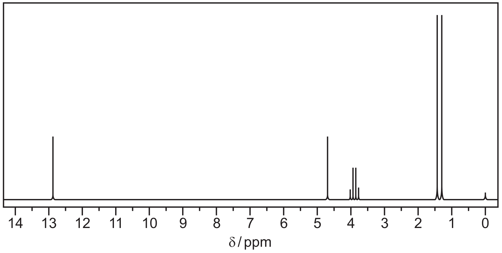
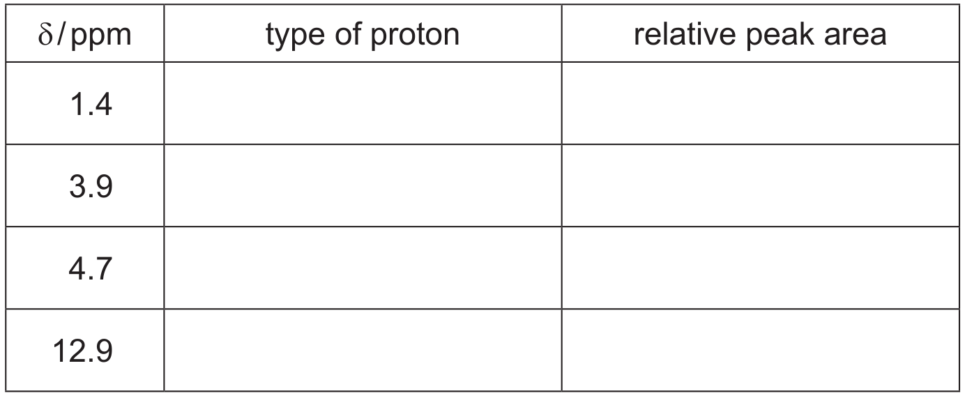
The proton NMR spectrum of M dissolved in shows eight peaks due to the eight different types of proton present in the molecule.
The proton NMR spectrum of M dissolved in was recorded.
Predict the number of peaks that would be seen in the proton NMR spectrum of M in . Explain your answer.
number of peaks explanation
[ 2 ]