EduNinja

IGCSE Biology2.77B ADH and urine concentrationTopic Practice

2.77B ADH and urine concentration

Understand why selective reabsorption of glucose occurs at the proximal convoluted tubule.

Question 1(d)(ii)

[Maximum number: 2]

Read the passage below. Use the information in the passage and your own knowledge to answer the questions that follow.

Treating Kidney Disease

Chronic kidney disease affects approximately 12% of the world's population and many people are unable to obtain treatment. One method of treating chronic kidney disease is by dialysis. The diagram shows how kidney dialysis is done.
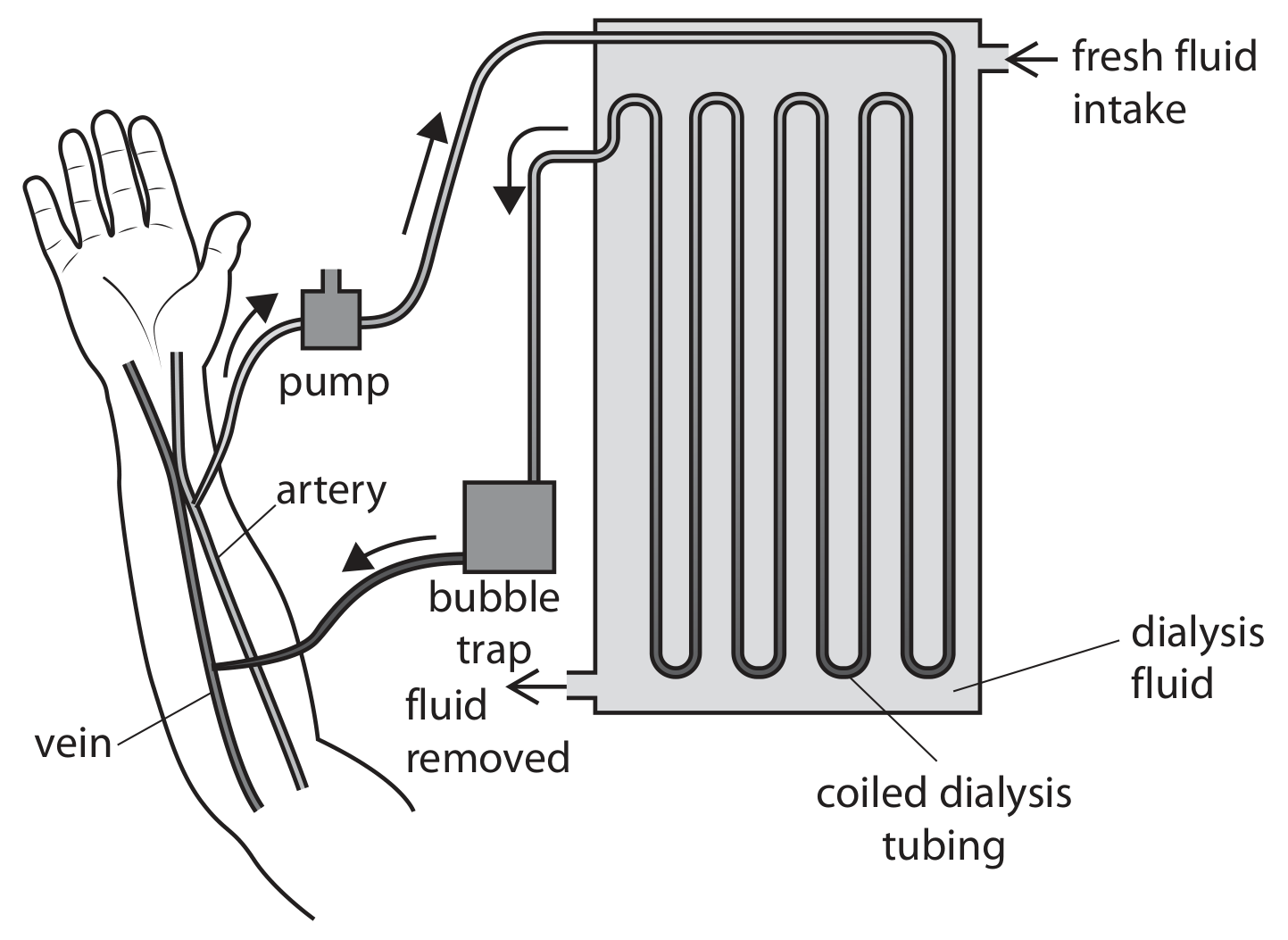
Question image

During dialysis, blood is taken from an artery in the arm and is pumped through a dialyser. In the dialyser, the blood passes through a long, coiled dialysis tube made of a thin partially permeable membrane. The tube is surrounded by dialysis fluid. The dialysis fluid contains glucose and ions at concentrations normally found in blood plasma, but does not contain urea. The urea passes from the blood into the dialysis fluid. The temperature in the dialyser is kept at 40C40^{\circ} \mathrm{C}. After passing through the dialyser, the blood is returned to a vein in the arm. Kidney dialysis can take up to three hours and must be done three times a week.

Scientists have now designed a new bioartificial kidney. This artificial kidney is a combination of engineering and living cells. This artificial kidney has a haemofilter made from artificial membranes that filter the blood. The substances filtered out of the blood then pass through a device called a bioreactor. This bioreactor absorbs useful substances back into the blood. The bioreactor is made of living nephron cells that are grown from stem cells. These cells are separated from the patient's blood by a silicon membrane to prevent the immune system rejecting them. Bioartificial kidneys are the same size as a human kidney and will be fitted inside the body to replace a kidney. The bioartificial kidney can react to changes in the body in the same way as a normal kidney.

The cells in the bioreactor absorb glucose from the filtrate in a way that is similar to the cells in a nephron (Lines 14 to 17).

Describe how the nephron absorbs glucose back into the blood from the filtrate.
Question 2(b)(i)

[Maximum number: 5]

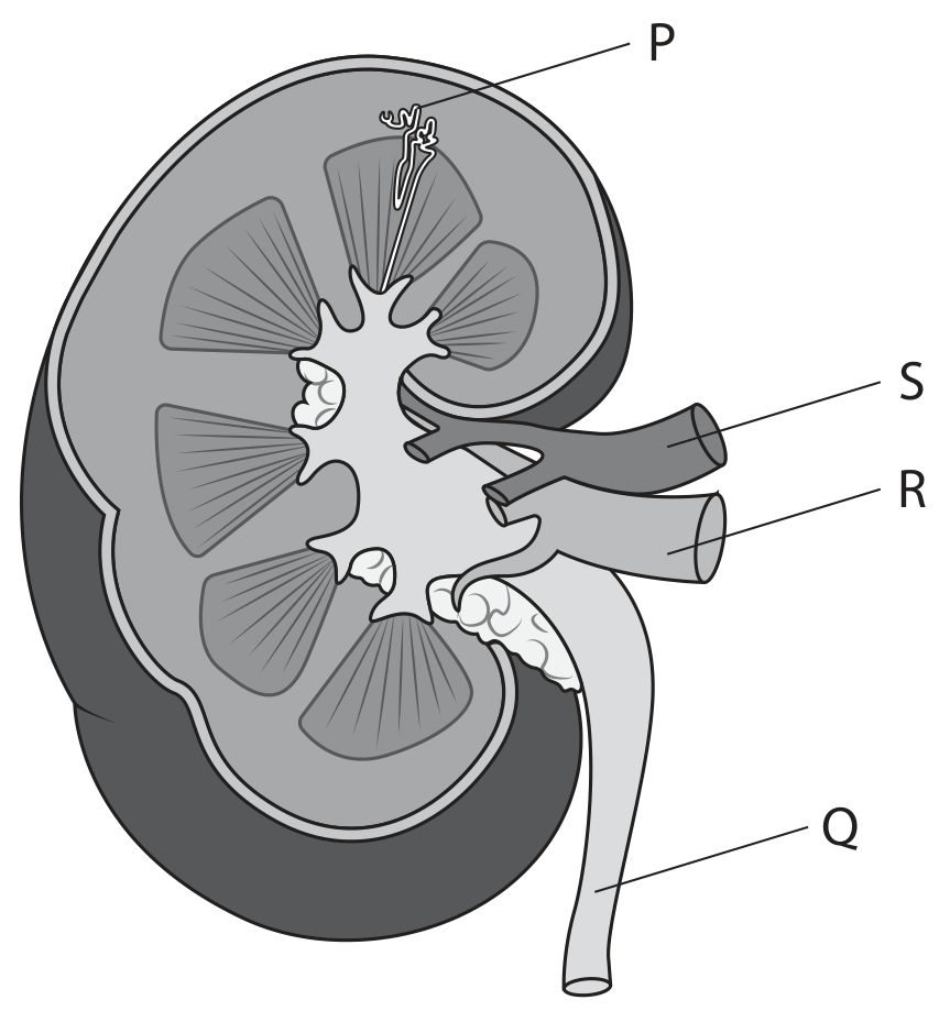
The diagram shows a section through a human kidney with some structures labelled.

Question image

Analysis of the composition of urine can be used in the diagnosis of problems with kidney function.

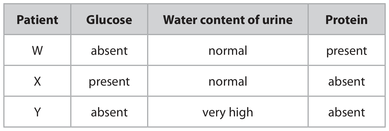
Urine samples are taken from three patients and the results are shown in the table.

Table

Discuss which processes in the kidney may be affected to produce the results shown.

Include in your answer where in the nephron the processes occur.

Question 2(a)(ii)

[Maximum number: 1]

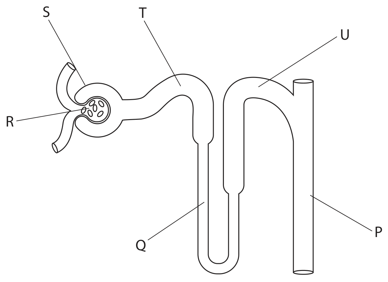
The diagram shows a nephron from a human kidney.

Question image

From which structure is glucose reabsorbed?

A

Q

B

R

C

S

D

U

Question 2(c)

[Maximum number: 4]

Mammals, such as humans, need to regulate their internal environment.

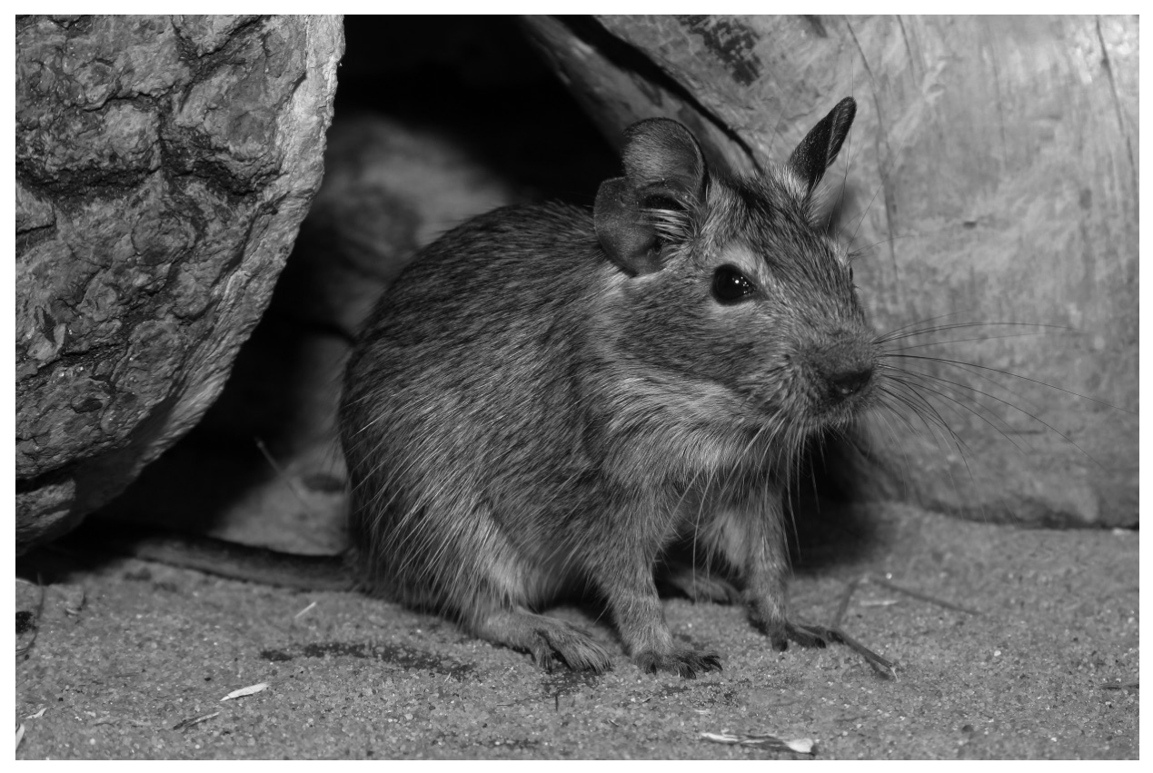
The photograph shows a mammal called a degu.

Question image

A degu is a rodent that lives in a very dry environment in South America. The degu gets its water input by feeding on plants.

The water intake of the degu changes during the winter and summer months.
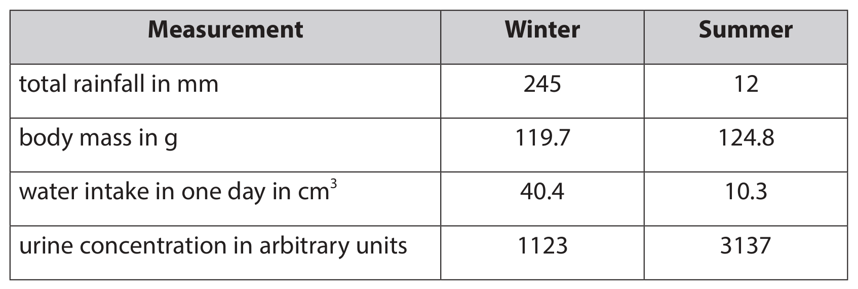
Table 2 shows the body mass, water intake and urine concentration of the degu in the winter and in the summer. It also shows the total rainfall in winter and in summer.

Table 2

Table 2

Comment on the differences in body mass, water intake and urine concentration in winter and summer.

Use the data in the table and your own knowledge in your answer.

Question 3(b)(i)

[Maximum number: 2]

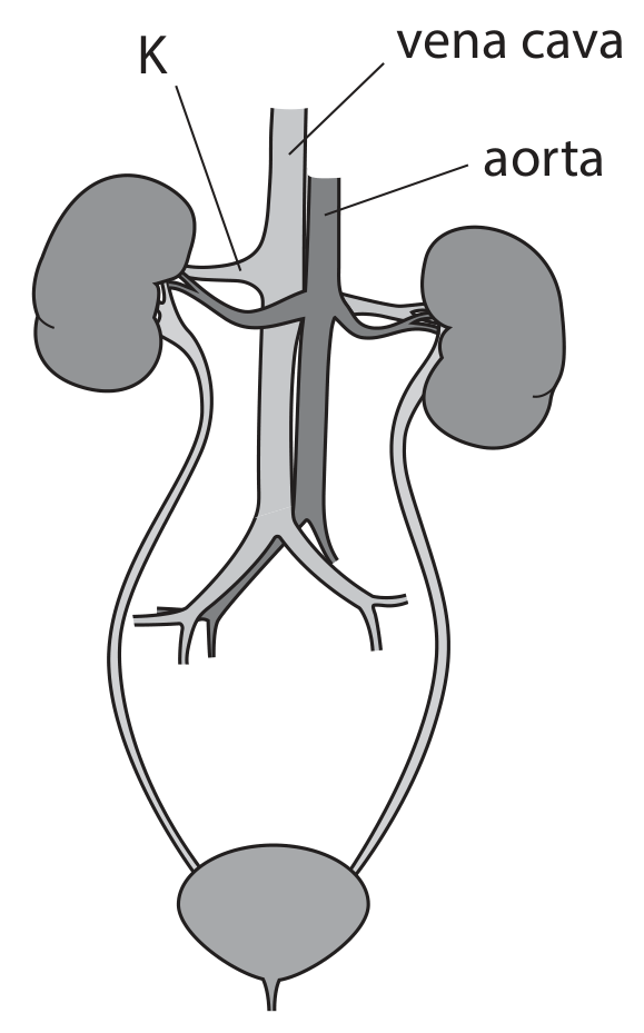
Diagram 1 shows the human urinary system with some structures labelled.

Diagram 1

Diagram 1

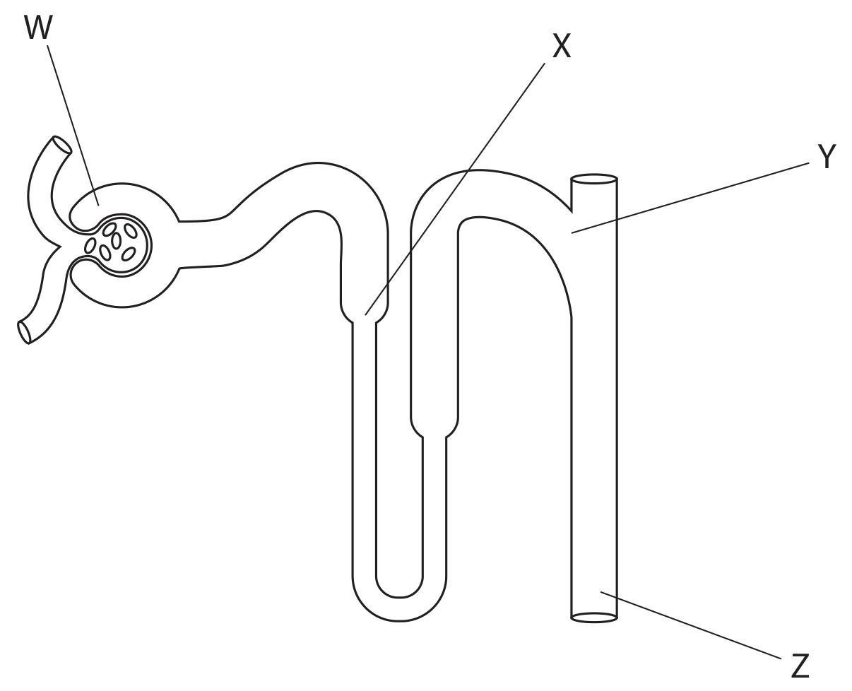
Diagram 2 shows a nephron with some parts labelled.

Diagram 2

Diagram 2

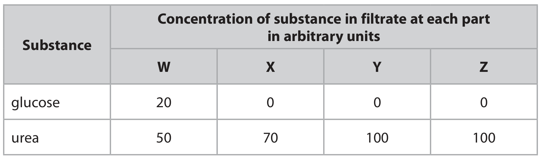
The table gives the concentrations of glucose and urea in the filtrate at parts W, X, Y and Z .

Table

Explain the difference in concentration of glucose between W and X .

Question 3(b)(i)

[Maximum number: 3]

The diagram shows a nephron from a human kidney with some structures labelled.

Question image

Blood plasma contains much glucose, but urine normally does not.

Explain what happens to glucose in the kidney.

Question 4(a)(ii)

[Maximum number: 1]

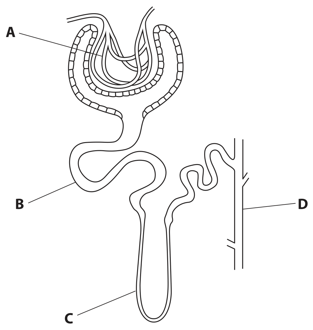
The diagram shows a nephron from a human kidney, with parts labelled A, B, C and D.

Question image

Use the diagram and your knowledge of kidney function to answer these questions.

Where does reabsorption of glucose take place?

Question 4(b)(ii)

[Maximum number: 2]

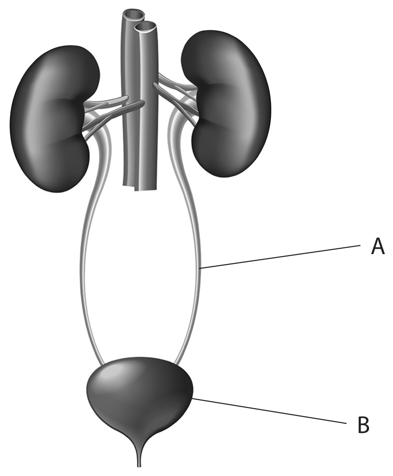
The diagram shows part of the human urinary system.

Question image

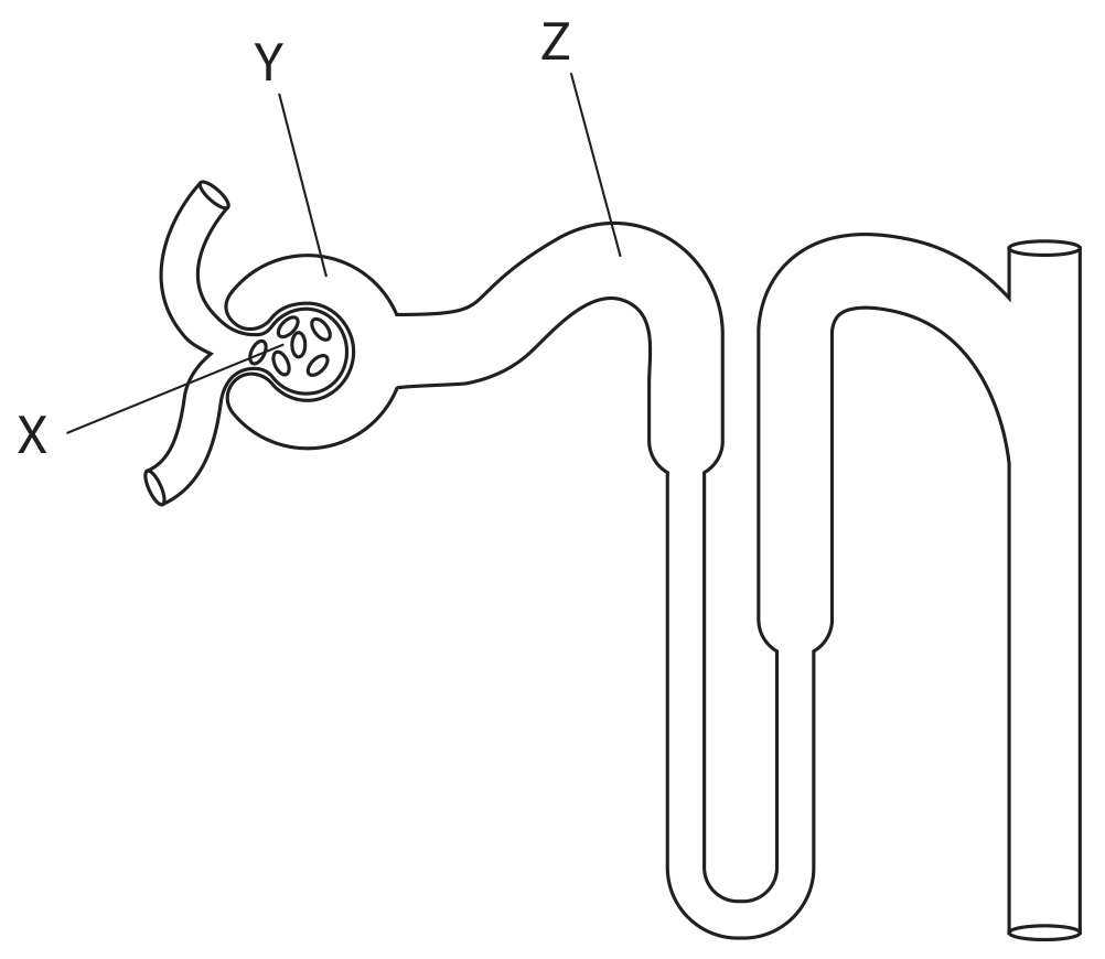
The diagram shows a kidney nephron.

Question image

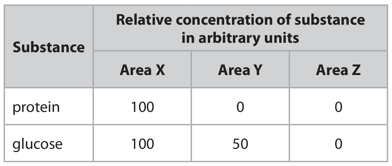
The table shows the relative concentrations of glucose and protein in the areas labelled X, Y and Z on the diagram.

Table

Explain the difference between the concentration of glucose in area Y and the concentration of glucose in area Z.

Question 5(a)(iv)

[Maximum number: 1]

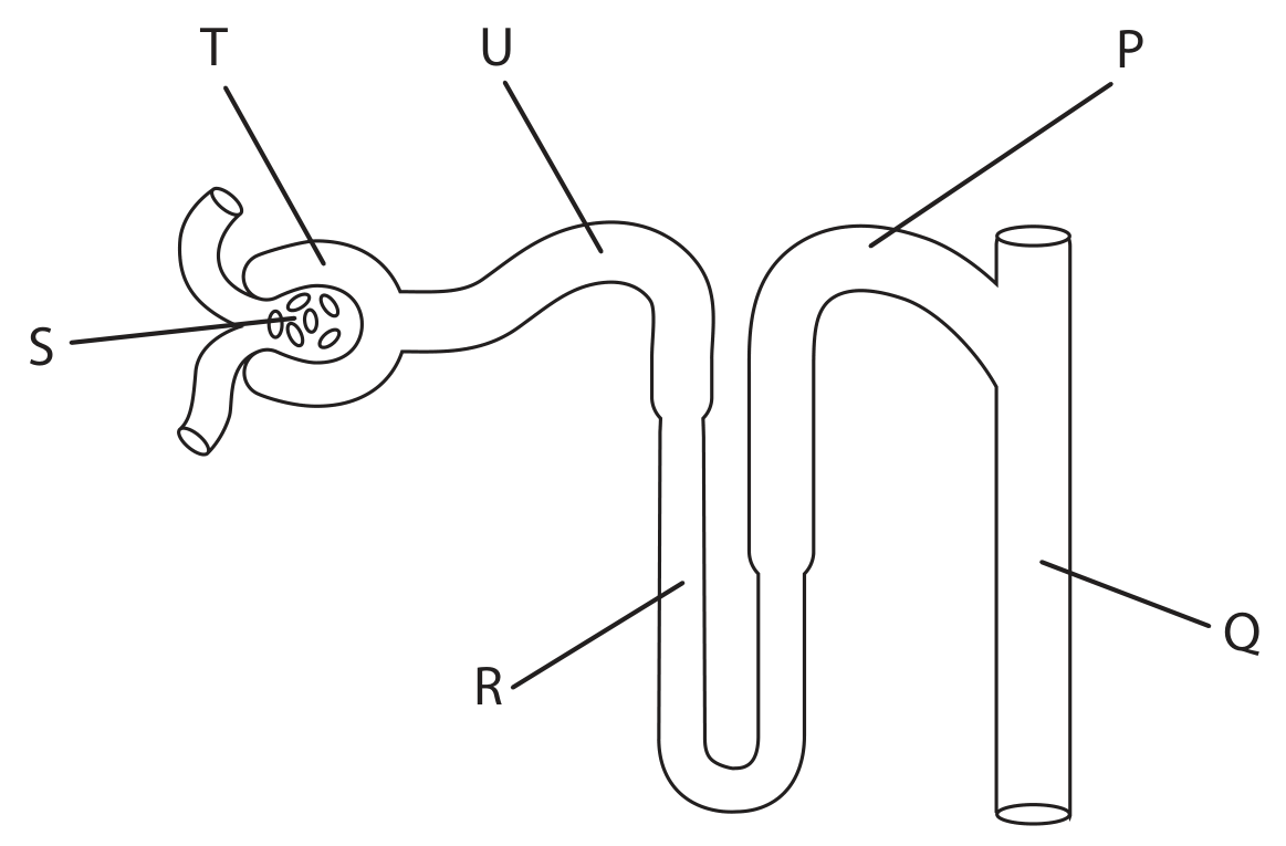
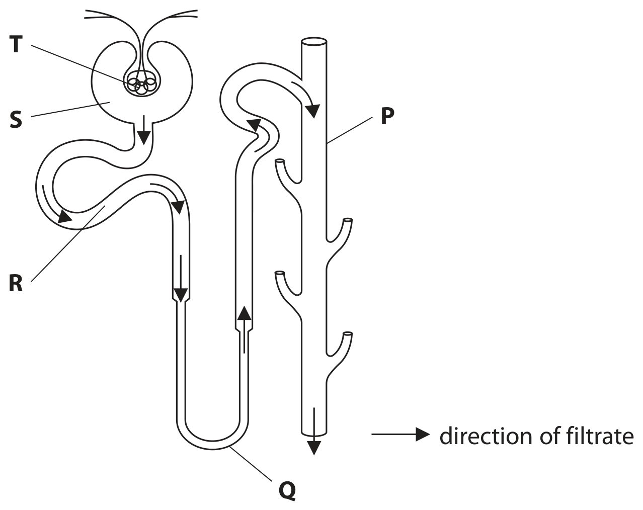
The diagram shows part of a human nephron, with parts labelled P, Q, R, S and T.

Question image

Where is glucose selectively reabsorbed into the blood?

A

Q

B

R

C

S

D

T

Question 5(a)

[Maximum number: 3]

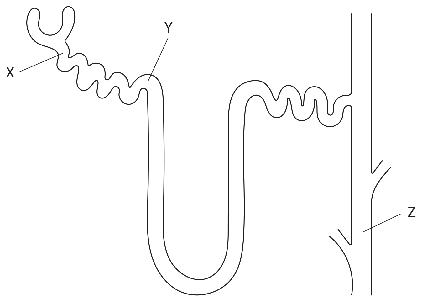
The diagram shows a nephron from a kidney with three different areas, X, Y and Z.

Question image

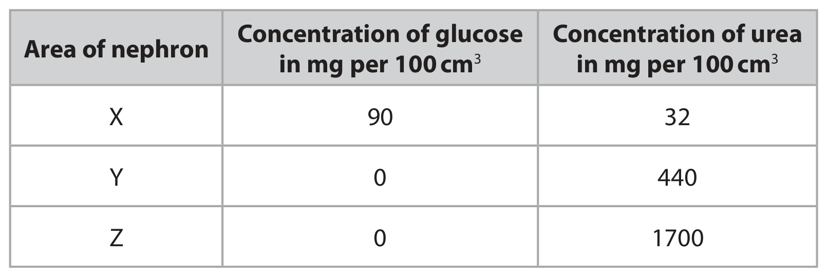
The table gives the concentration of glucose and urea at X, Y and Z.

Table

Explain the difference in concentration of glucose between X and Y .

0 selected