Question 1
[Maximum number: 2]
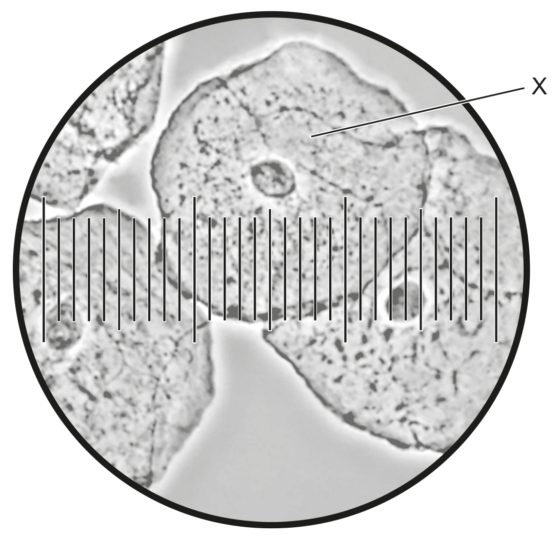
The micrograph is an image of human cheek cells viewed with a light microscope.
Question 1(c)
(a)
The electron micrograph shows part of an animal cell.
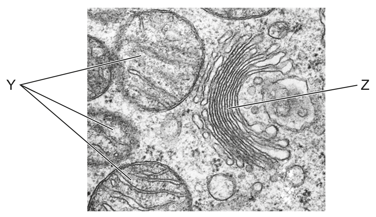
Outline the functions of Y and Z in the cell.
[ 2 ]
 EduNinja
The micrograph is an image of human cheek cells viewed with a light microscope.
The electron micrograph shows part of an animal cell.
Outline the functions of Y and Z in the cell.
The image shows an electron micrograph of pancreatic exocrine cells.
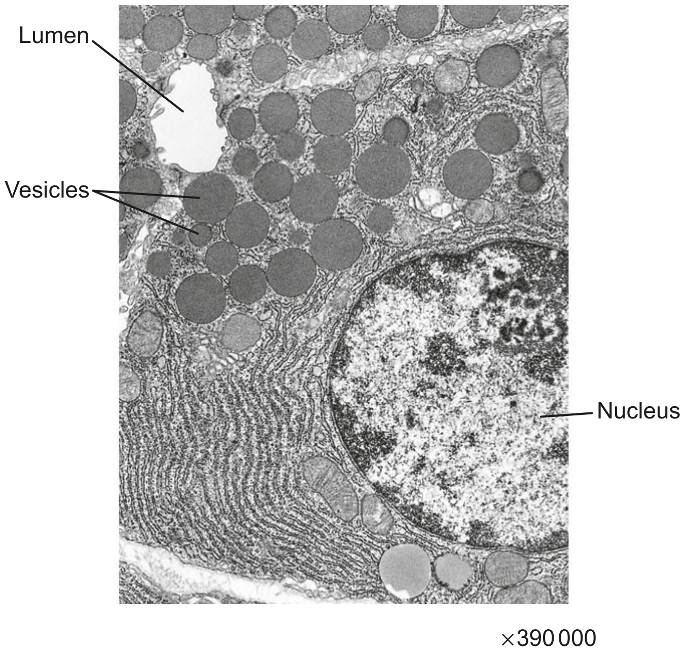
What is the role of the vesicles shown in the micrograph?
To transport hormones between the rough endoplasmic reticulum and the Golgi apparatus
To store glycogen when blood glucose levels are high
To move enzymes out of the cell by exocytosis
To digest cellulose
Where are proteins synthesized by free ribosomes used?
Outside the cell after secretion
Within the nucleus
Within the lysosomes
Within the cytoplasm
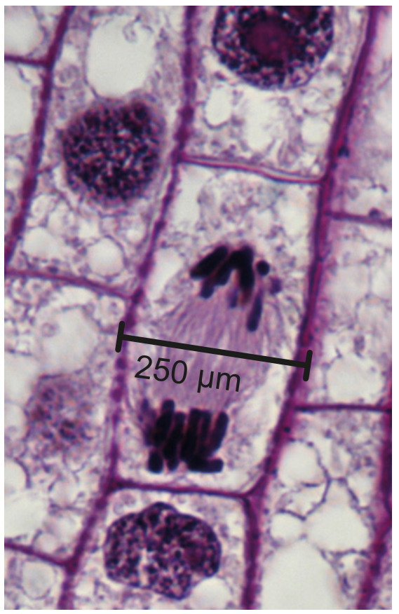
The micrograph shows a cell from the root of an onion (Allium cepa) during mitosis.
State what is indicated by the presence of polysomes in a cell.
In which image are polysomes visible? (The images do not have the same magnification)
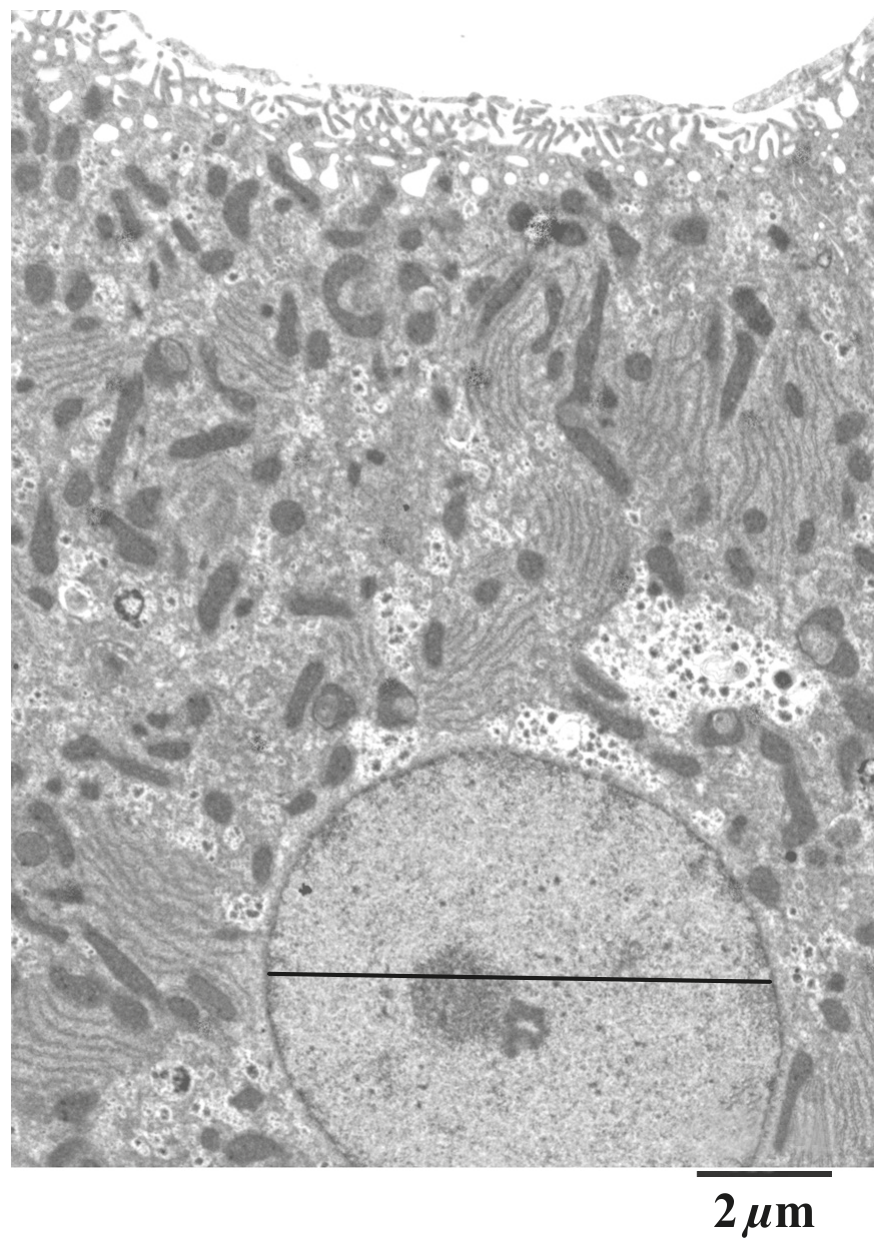
The electron micrograph shows part of a pancreas cell that secretes digestive enzymes.
State the organelle where digestive enzymes are synthesised.
The following shows an electron micrograph of a liver cell.
The electron micrograph is a higher magnification of a liver cell.
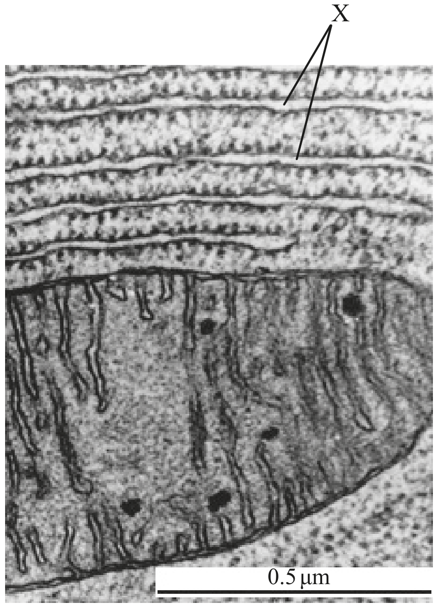
State its main function.
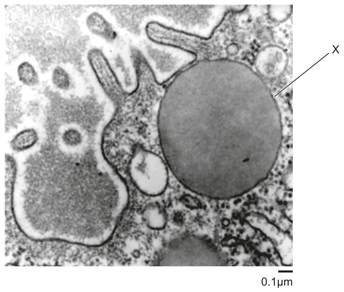
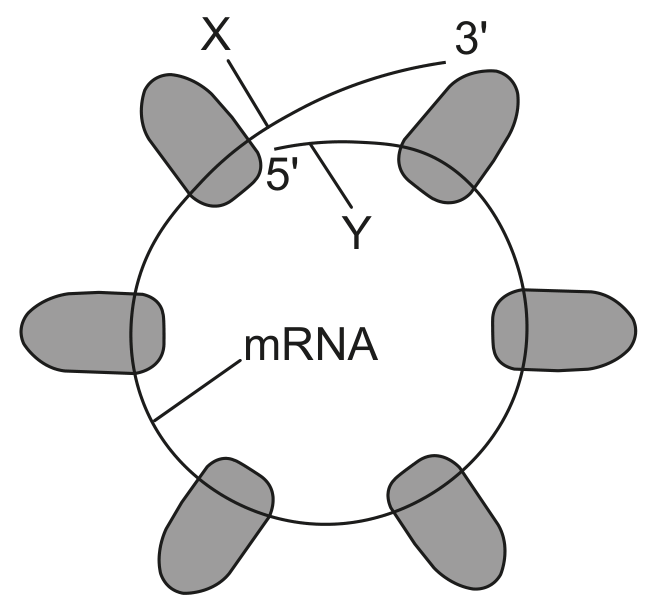
The electron micrograph shows part of a cell in the pituitary gland of a rat. Ribosomes appear as dark granules. Some of the ribosomes are arranged in a linear array and some are in circles. The diagram shows how the ribosomes in a circular array are connected.
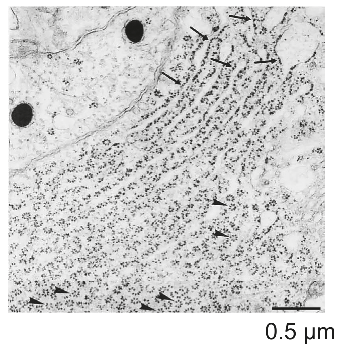
On the electron micrograph, label
On the electron micrograph, label the rough endoplasmic reticulum.
On the electron micrograph, label a polysome.
The diagram shows how vesicles are used to transport materials in a cell.
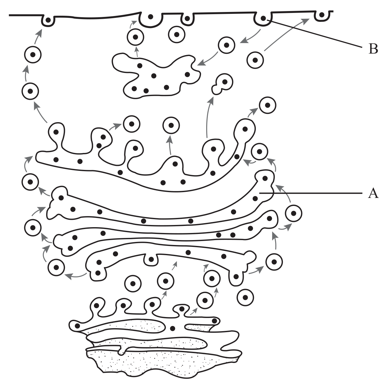
State the name of organelle A .
Outline what happens to the proteins produced by free and bound ribosomes.
Free ribosomes:
Bound ribosomes: