Question 1
[Maximum number: 1]
Which function is accomplished by structures X and Y in the Paramecium?
 EduNinja
Which function is accomplished by structures X and Y in the Paramecium?
Which feature of striated muscle cells allows them to be considered as a possible exception to the cell theory?
They are found in multicellular organisms.
They contain more than one nucleus.
They are specialized for movement.
They do not carry out mitosis.
What cell component is found in eukaryotic cells but not in prokaryotic cells?
Mitochondria for respiration
DNA containing genetic information
Ribosomes for protein synthesis
Cell wall to maintain shape
The magnification of the micrograph is .
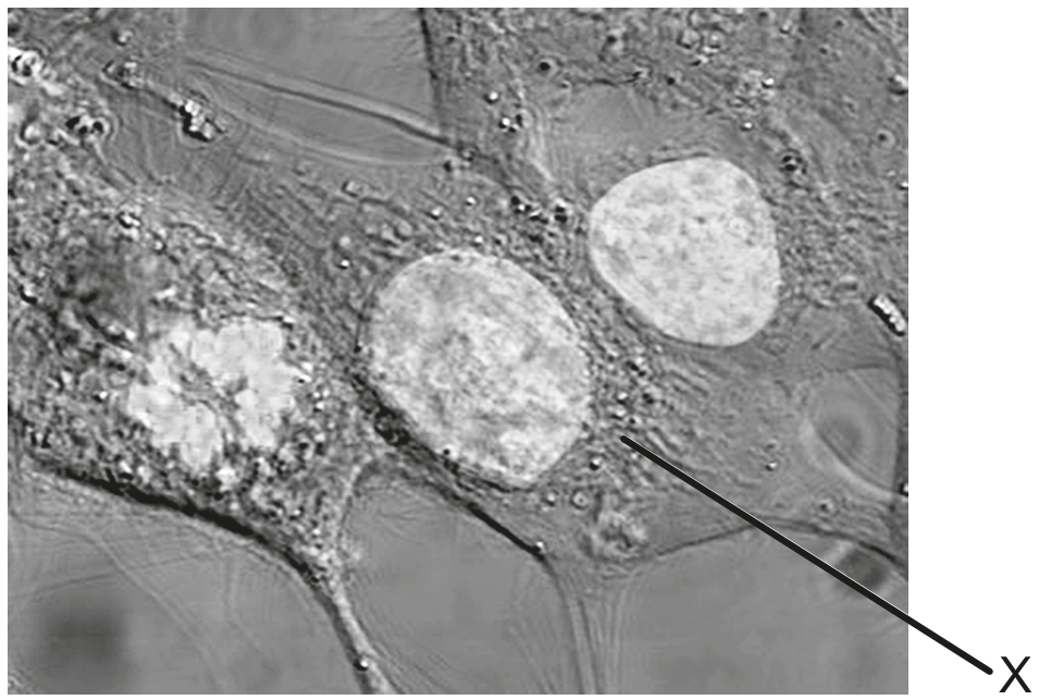
What is the maximum diameter of the nucleus in the cell labelled X ?
10 nm
20 nm
Chlorella and Paramecium are both unicellular eukaryotic organisms living in freshwater. Chlorella is photosynthetic and has a cell wall. Which organelle will be found in Paramecium but not in Chlorella?
Chloroplast
Contractile vacuole
Rough endoplasmic reticulum
Mitochondrion
In mammals, mature red blood cells are specialized in that they lack nuclei, mitochondria or ribosomes. Which statement applies to red blood cells?
No chemical reactions take place within their cytoplasm.
They cannot produce new enzymes.
Materials cannot enter red blood cells.
Materials cannot exit red blood cells.
The images of the radiolarian, a single-celled marine organism, were produced using a light microscope (left) and a scanning electron microscope (right).
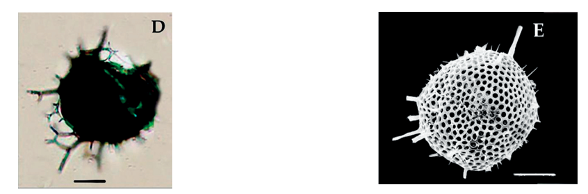
What is a reason for the difference in quality of these images?
Light cannot pass through the specimen.
Higher magnification can be achieved with the electron microscope.
The resolution of the electron microscope is higher.
Samples are stained with methylene blue when viewed with the light microscope.
The micrograph is an image of human cheek cells viewed with a light microscope.
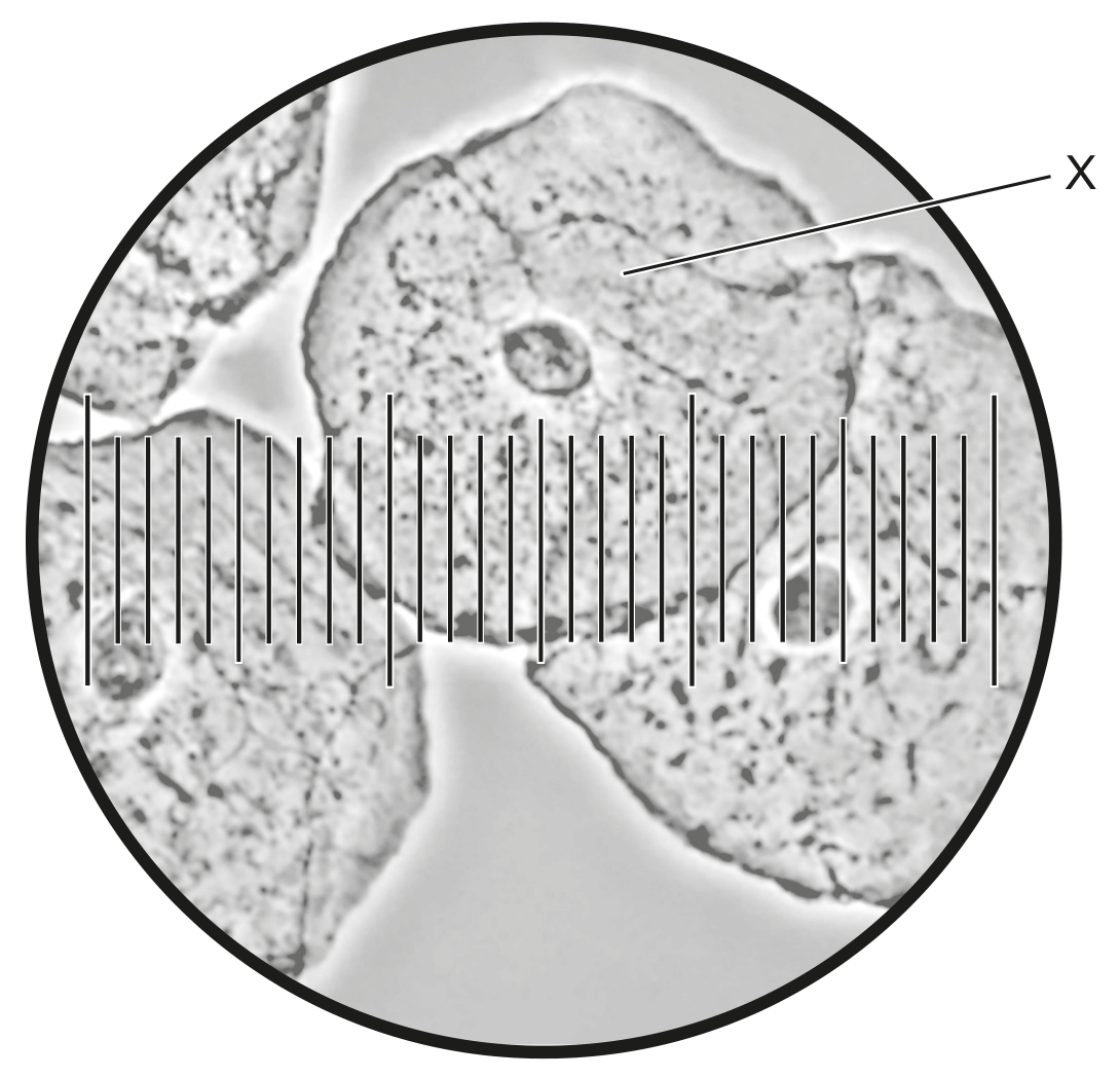
Identify the piece of equipment that is used together with the microscope to measure the size of the cells.
Each small division of the scale in the micrograph is equivalent to .
Calculate the diameter of the cell labelled X .
Calculate the magnification of the image.
The micrograph shows two types of blood cell.
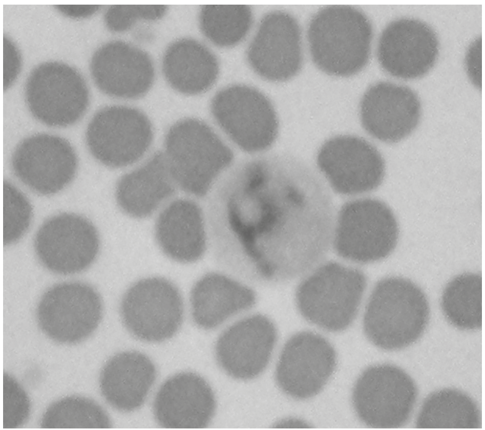
What determines the differences between the two types of cell?
Different number of mitotic cycles
Different expression of some genes
Reaction to oxygen of red blood cells
Reaction to antigens of white blood cells
The photomicrograph shows a section of onion (Allium cepa) root tip cells.
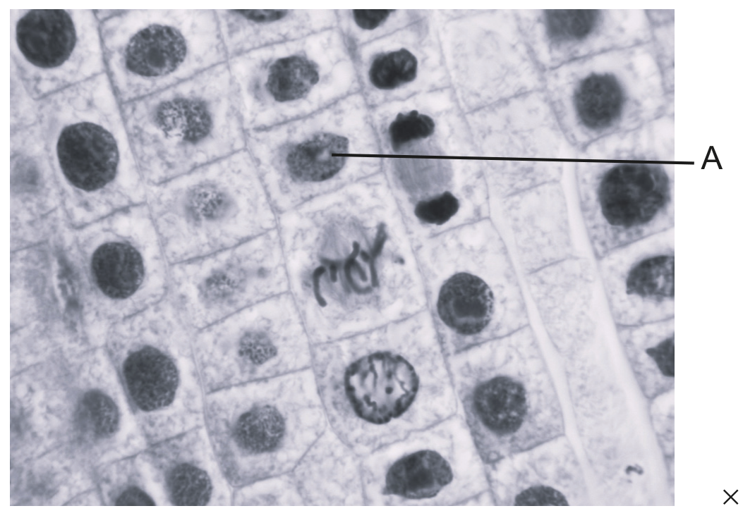
× 1000
Calculate the actual length of the cell labelled A , giving the units.