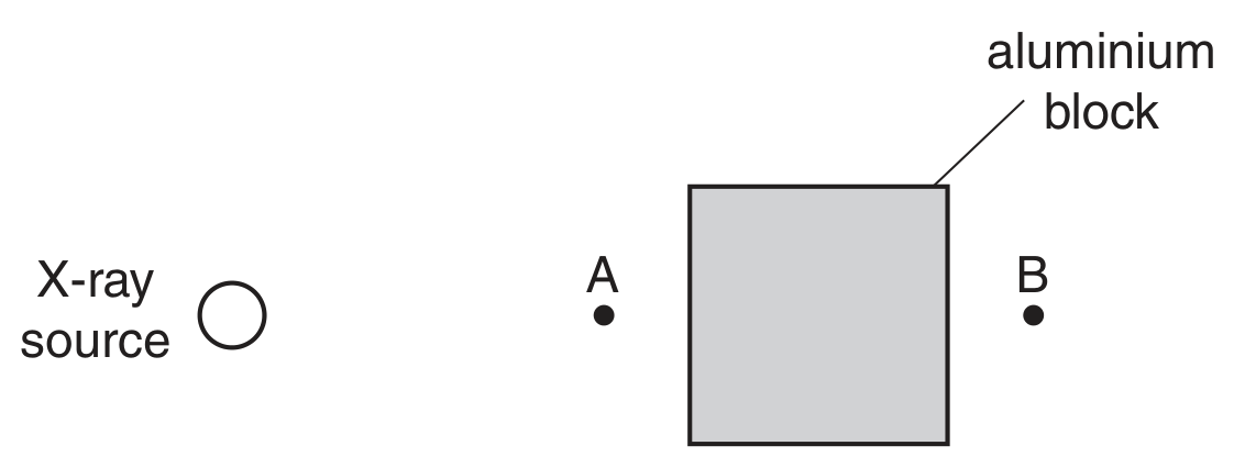
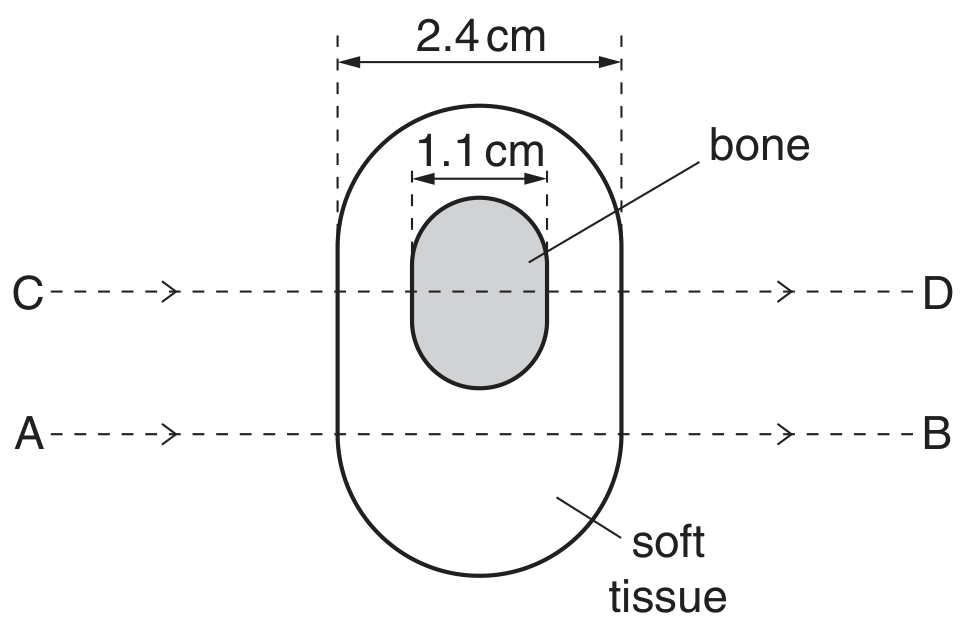
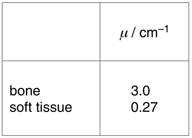
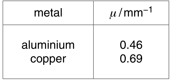
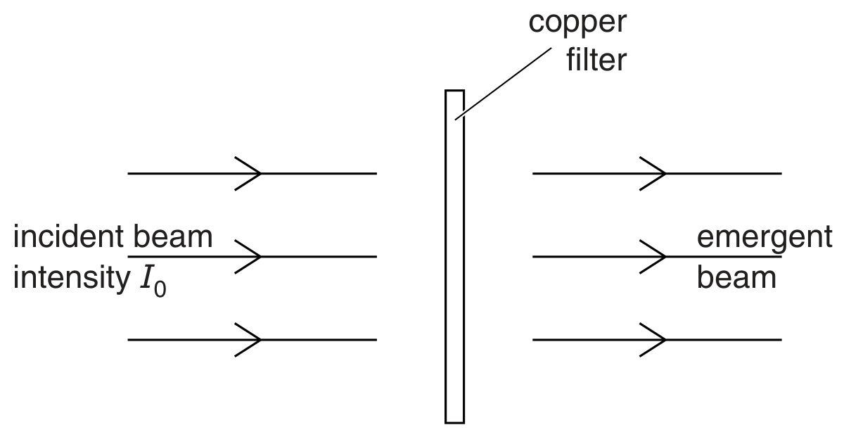
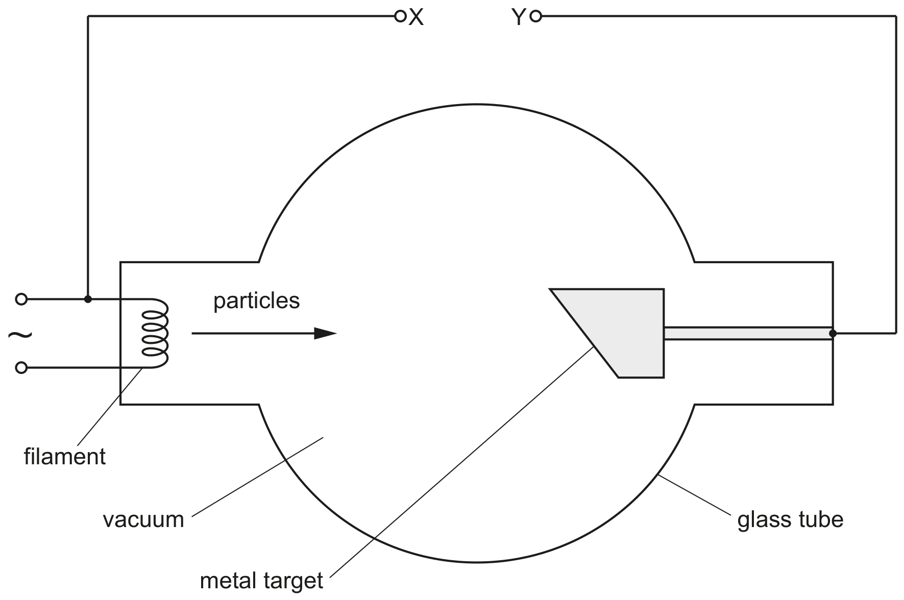
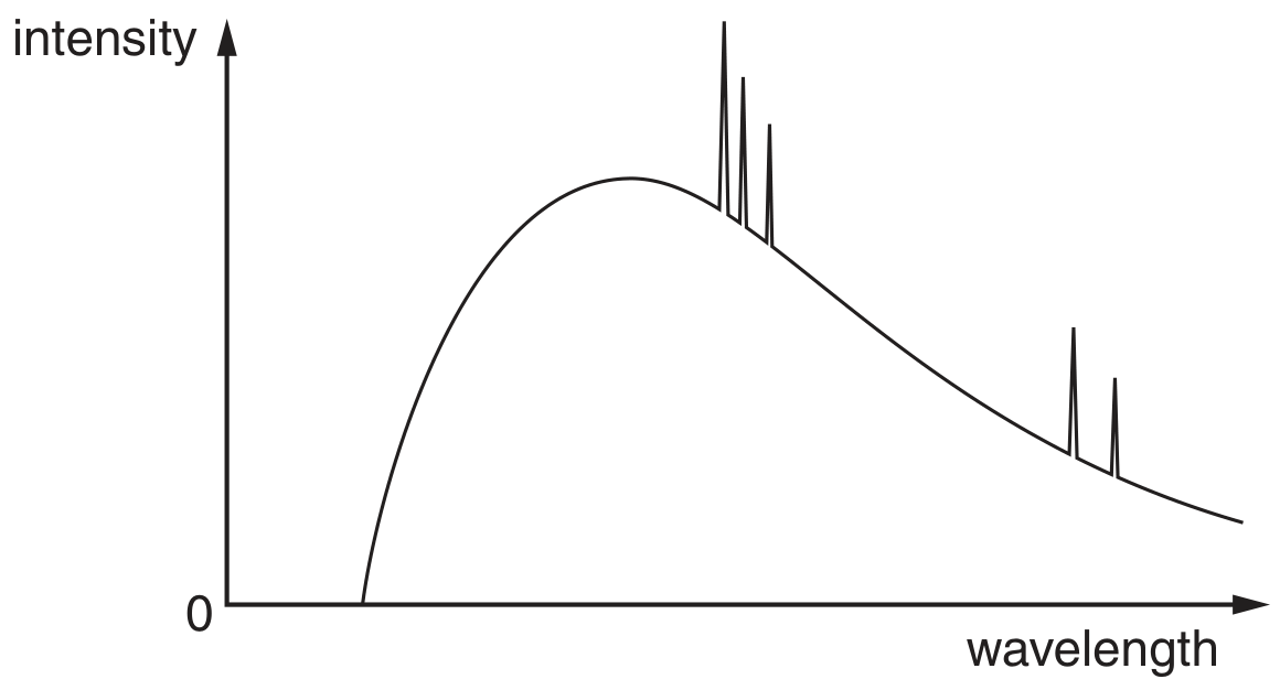
Question 7
Question 7(b)
(a)
Four electron energy levels in an atom are shown in Fig. 7.1.
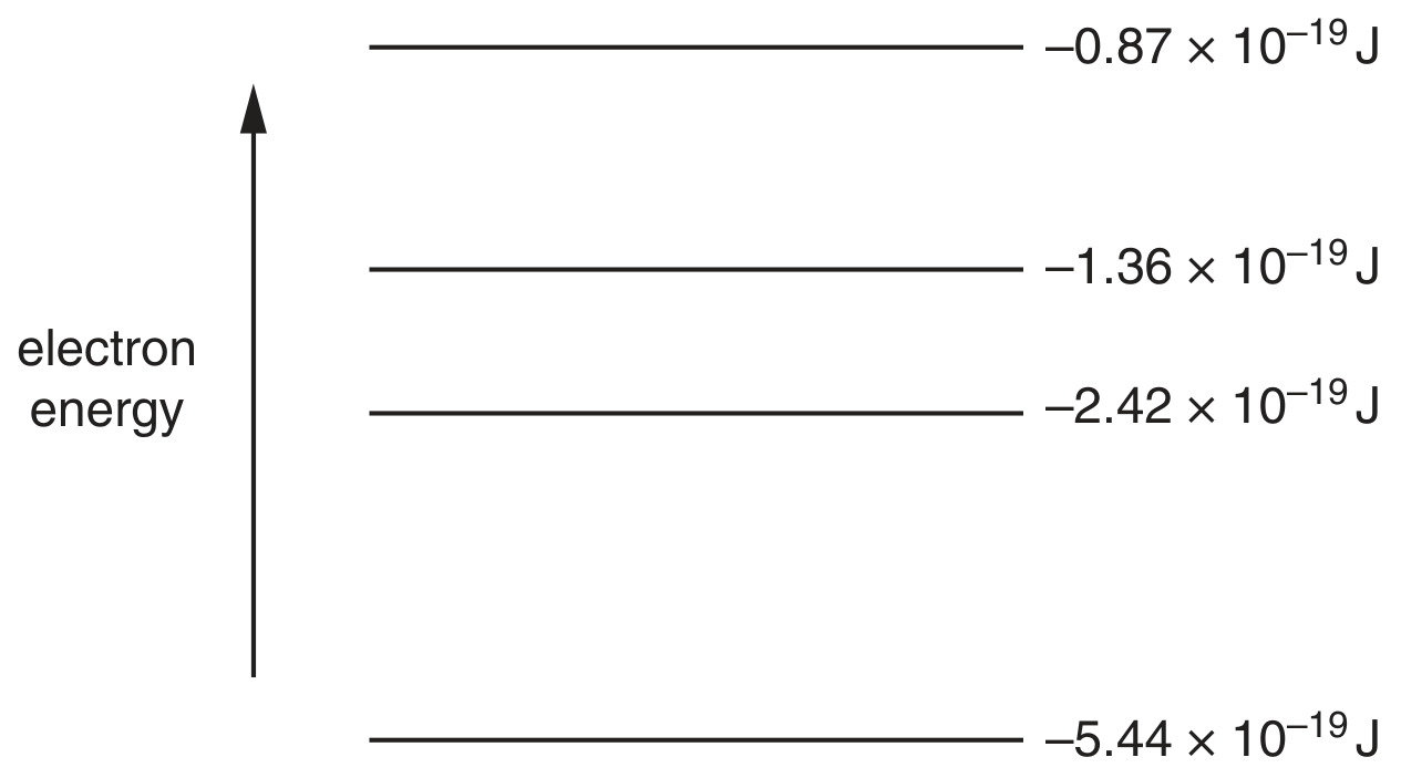
Fig. 7.1 (not to scale)
An emission spectrum is associated with the electron transitions between these energy levels.
For this spectrum,
[ 2 ]
Question 7(b)(ii)
(i)
calculate the minimum wavelength.
wavelength = m
[ 2 ]