Q BankQuestion BankDocsDocuments

B2.1 Membranes and membrane transport

Membranes and membrane transport link bilayer structure, membrane proteins, selective permeability, gradients, and vesicle movement to cellular control; Students connect membrane structure to transport mechanisms across cellular boundaries.

Syllabus
First assessment 2025
Topic
B2.1
Level
HL

Bilayers Put a Water-Resistant Core Between Compartments

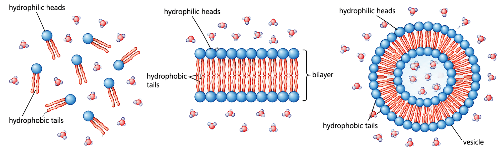
A lipid bilayer forms because amphipathic phospholipids place water-compatible heads toward water and hydrocarbon tails away from it.

In water, this arrangement lowers unfavorable tail–water contact. The two leaflets therefore create a flexible sheet with a non-polar interior, allowing a cell to separate its inside from its surroundings.

A membrane has water on both sides: heads face each aqueous side while tails meet in the middle, forming one continuous barrier.

When phospholipids are placed in water, their hydrophilic heads remain in contact with water while their hydrophobic tails cluster away from it; a closed bilayer can therefore form without leaving exposed tails at its edge.

The bilayer is a consequence of amphipathic structure in an aqueous environment; it is not a rigid wall, and its hydrophobic core still permits some small non-polar molecules to cross.

Lipid bilayers as basis

Assessment in practice

1–3 marks
How it is assessed

This objective is assessed through structured response, commonly using Identify / Explain / State.

Command terms

Identify / Explain / State / Outline / Draw / Label

What earns marks

Build the answer around this relationship: Phospholipids have hydrophilic phosphate heads and hydrophobic fatty acid tails.

Watch for

Saying phospholipids are only hydrophobic or only hydrophilic instead of amphipathic.

Representative question

Question 1

[Maximum number: 8]

Cell membranes separate aqueous environments in cells. Explain how the properties of phospholipids help to maintain the structure of cell membranes.

The Bilayer Is a Selective Barrier

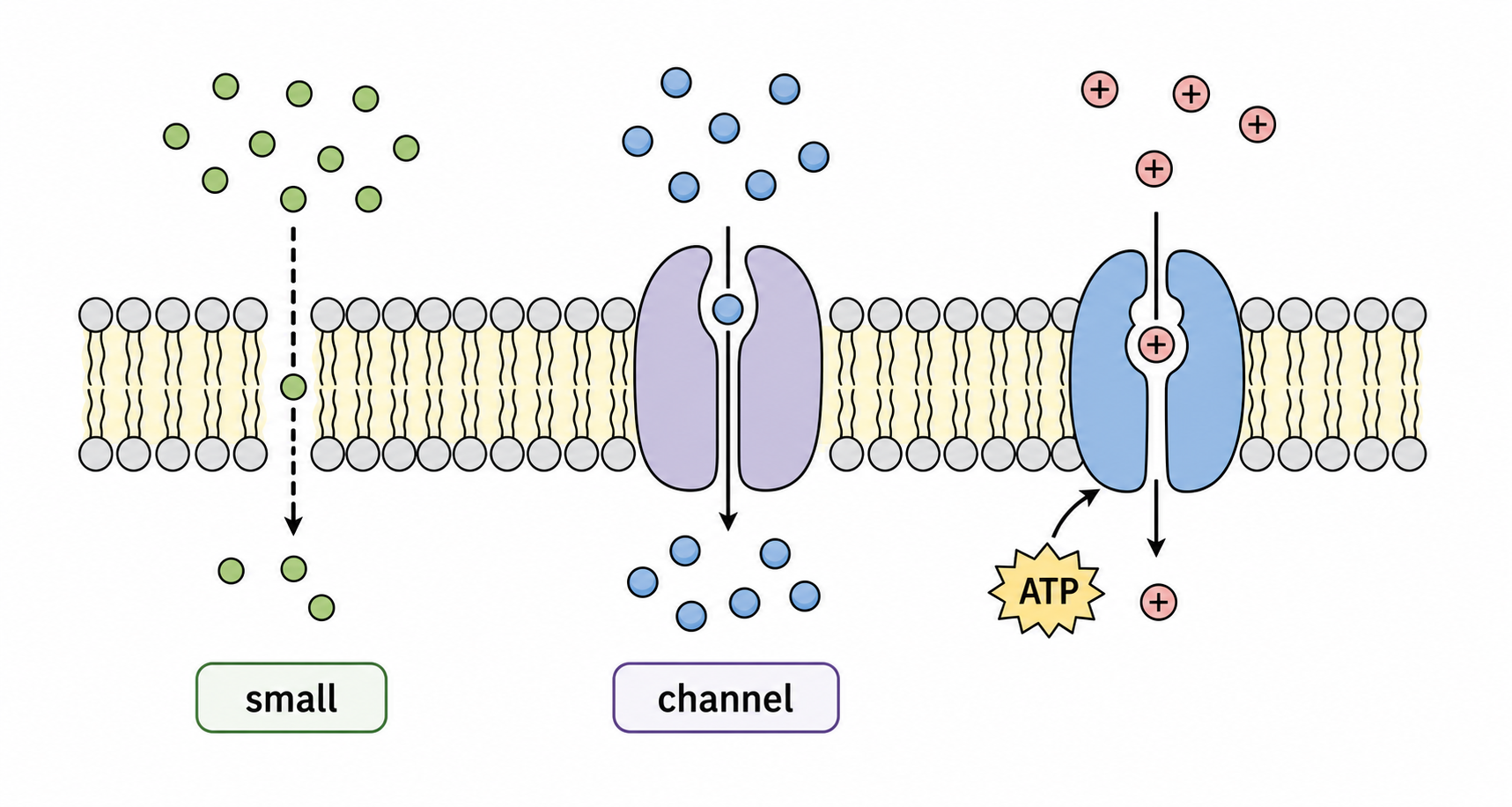
The bilayer’s hydrophobic interior slows ions and most polar molecules, while small non-polar molecules cross more readily.

Crossing depends on how well a solute interacts with the tail region and on its size. The membrane can therefore separate concentration gradients without blocking every substance equally.

Use three checks: charge/polarity, size, and whether a transport protein is available. A matching pathway can change the prediction.

Oxygen can diffuse through the tail core, but a sodium ion needs a channel or carrier because its charge is incompatible with the hydrophobic interior.

‘Barrier’ does not mean ‘impermeable’. Selectivity comes from different crossing rates, not from stopping all movement.

Bilayers as barriers

Assessment in practice

1 marks
How it is assessed

This objective is assessed through multiple choice, commonly using Identify.

Command terms

Identify

What earns marks

Build the answer around this relationship: The bilayer core is hydrophobic because fatty acid tails point inward.

Watch for

Treating polar glucose as able to pass directly through the phospholipid core.

Representative question

Question 1

[Maximum number: 1]

Neural pathways in living brains can now be mapped by tracking the movement of water molecules inside axons. What keeps water molecules inside axons?

A

Plasma membrane

B

Hydrogen bonding

C

Pump proteins

D

Synapse

Simple Diffusion Runs Down a Gradient

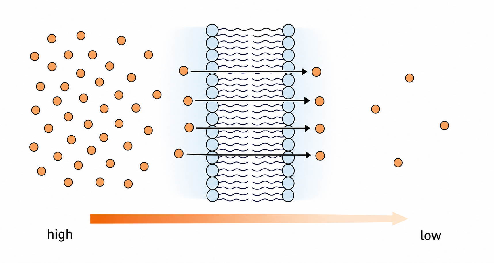
Simple diffusion is net movement from higher concentration to lower concentration directly through the membrane, without ATP or a transport protein.

Random molecular motion is present in both directions, but more particles leave the high-concentration side, producing net movement down the gradient until equilibrium is approached.

Check: concentration gradient exists; molecule can cross the lipid core; no energy input or protein is required.

If oxygen is higher outside a cell than inside, oxygen molecules cross in both directions but net oxygen entry occurs until the gradient shrinks.

Diffusion is not movement toward ‘where the cell needs it’. The gradient and membrane permeability determine net direction.

Simple diffusion

Assessment in practice

1–3 marks
How it is assessed

This objective is assessed through structured response, commonly using Compare / Outline / Explain.

Command terms

Compare / Outline / Explain / Identify

What earns marks

Build the answer around this relationship: Simple diffusion is passive movement from higher to lower concentration.

Watch for

Adding channel proteins or ATP to simple diffusion when the row requires direct passive movement.

Representative question

Question 1

[Maximum number: 3]

Explain how the rate of diffusion of oxygen into a cell is affected by the concentration of oxygen outside of the cell.

Membrane Proteins Sit in Different Places

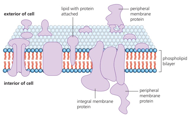
Integral membrane proteins are embedded in or span the bilayer; peripheral proteins attach to a membrane surface or to another protein.

Hydrophobic amino-acid regions can sit among lipid tails, while polar regions face water or form pathways. Surface attachment does not require a protein to cross the hydrophobic core.

Classify by location: embedded/spanning means integral; surface-associated without entering the core means peripheral. Then connect location to likely function.

A channel crossing the membrane is integral; a cytoskeletal protein attached to its cytoplasmic face is peripheral.

A protein being near a membrane does not prove it spans the bilayer. Identify whether it enters the hydrophobic core.

Integral and peripheral proteins

Assessment in practice

1–4 marks
How it is assessed

This objective is assessed through structured response, commonly using State / Identify / Outline.

Command terms

State / Identify / Outline / Describe / Explain / Label

What earns marks

Build the answer around this relationship: Integral proteins are embedded in the bilayer and may span it completely.

Watch for

Listing non-membrane processes such as DNA replication or glycolysis as membrane protein functions.

Representative question

Question 1

[Maximum number: 4]

Describe the functions of proteins in cell membranes.

Osmosis Is Water Movement Through a Selective Membrane

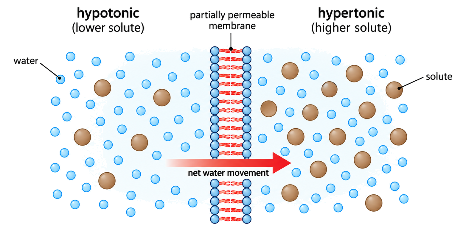
Osmosis is net movement of water across a partially permeable membrane from higher water potential to lower water potential.

Solute lowers the water potential of a solution. Water molecules move randomly both ways, but the imbalance in water potential produces net movement toward the more concentrated solution; aquaporins can increase the rate.

State the membrane and compare water potential: higher ψ → lower ψ; then predict volume or cell-size change.

A cell placed in a solution with lower water potential loses water through its membrane and may shrink; aquaporins change speed, not the direction set by ψ.

Osmosis is not simply ‘water moves to higher solute’ without conditions: the membrane must allow water through and the water-potential comparison must be valid.

Osmosis and aquaporins

Assessment in practice

1–2 marks
How it is assessed

This objective is assessed through structured response, commonly using Describe / Identify / Define.

Command terms

Describe / Identify / Define / State / Explain / Distinguish

What earns marks

Build the answer around this relationship: Osmosis is passive net movement of water across a selectively permeable membrane.

Watch for

Describing movement of solute instead of net movement of water.

Representative question

Question 1

[Maximum number: 4]

Describe transport across cell membranes by osmosis.

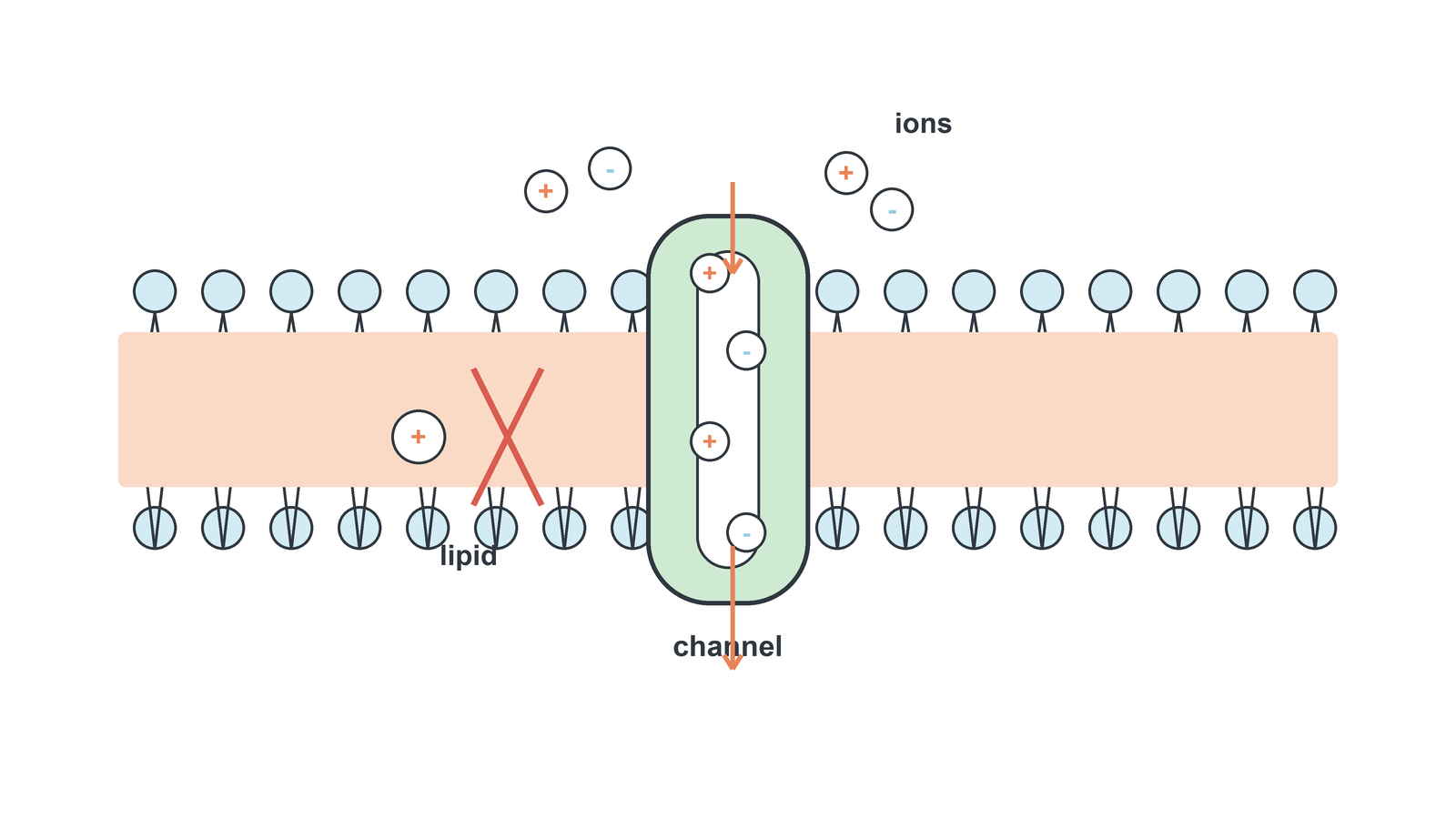
Channels Give Certain Solutes a Hydrophilic Route

Channel proteins form selective hydrophilic pores through the bilayer, allowing particular ions or polar molecules to cross down an electrochemical gradient.

The pore exposes a compatible interior while the surrounding lipid tails remain excluded. Size, charge and binding sites determine which solutes pass; channels do not normally supply the energy for uphill movement.

For a channel claim, name the solute, pore selectivity and gradient direction.

An ion channel can let potassium move rapidly down its gradient while excluding a larger or differently charged ion.

A channel is not automatically an active pump. If movement is against a gradient, another energy-coupled mechanism is required.

Channel proteins

Assessment in practice

1 marks
How it is assessed

This objective is assessed through structured response, commonly using Identify / Explain / Describe.

Command terms

Identify / Explain / Describe / Outline

What earns marks

Build the answer around this relationship: Channel proteins form hydrophilic pathways through the membrane.

Watch for

Adding ATP to facilitated diffusion even when the movement is passive.

Representative question

Question 1

[Maximum number: 3]

Explain facilitated diffusion.

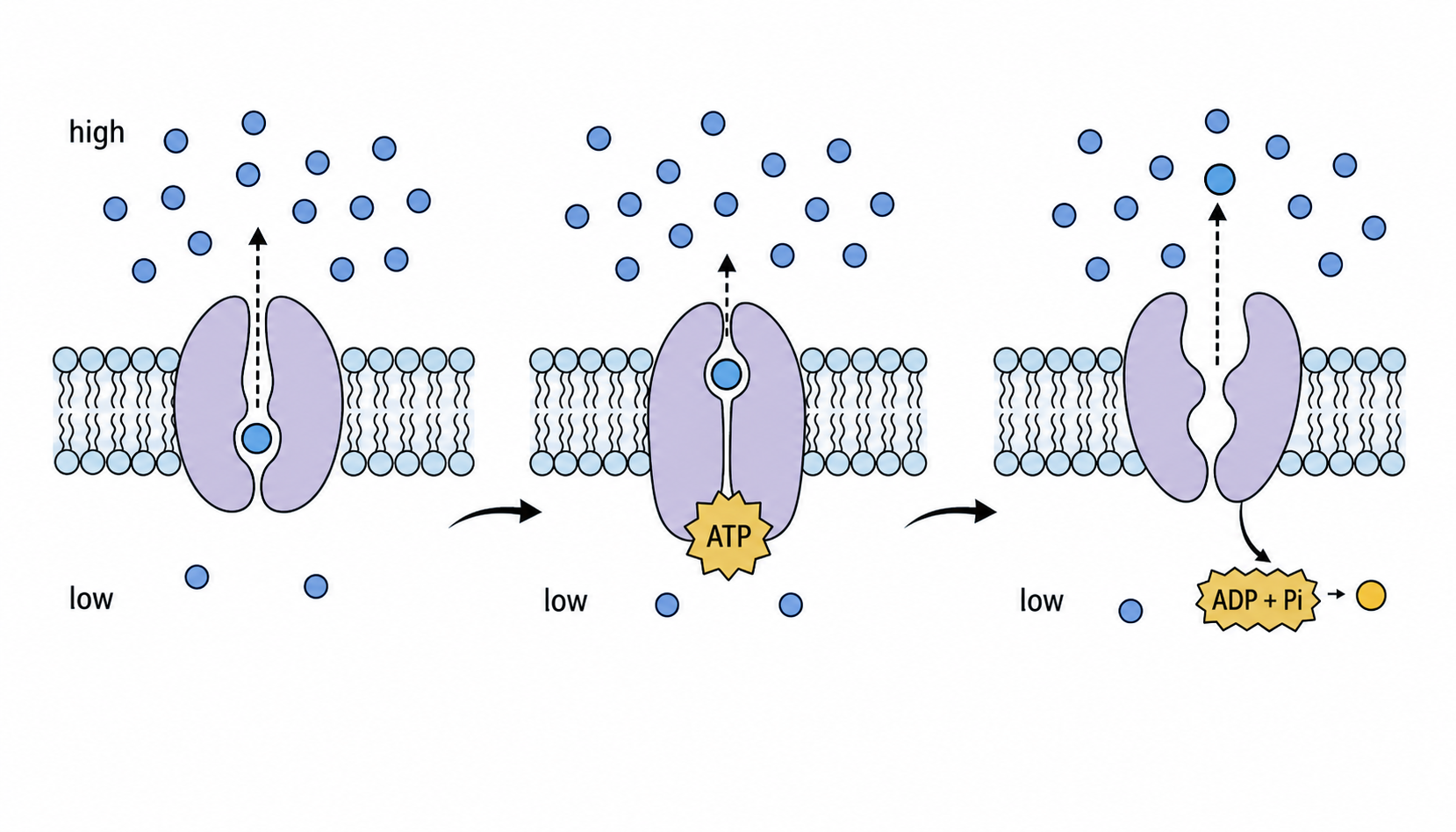
Pump Proteins Use Energy to Move Uphill

Pump proteins use energy, commonly from ATP hydrolysis, to move selected substances against their concentration or electrochemical gradient.

A pump changes conformation when energy is supplied, alternately exposing a binding site to each side of the membrane. This couples an unfavorable transport step to a favorable energy-releasing reaction.

Trace: bind solute → energy changes protein shape → release solute on the opposite side → reset.

An ATP-driven pump can keep sodium higher outside a cell even though diffusion would tend to move sodium inward.

A protein moving a solute is not enough to identify active transport; look for uphill movement and an energy source.

Pump proteins

Assessment in practice

1–3 marks
How it is assessed

This objective is assessed through structured response, commonly using Explain / Identify / Deduce.

Command terms

Explain / Identify / Deduce

What earns marks

Build the answer around this relationship: Pump proteins use ATP to move substances across membranes.

Watch for

Calling pump proteins channels when the mark scheme rejects channels for active transport.

Representative question

Question 1

[Maximum number: 3]

Calcium is absorbed from food in the human gut by both active and passive processes. Outline active transport, including the benefits of the process.

Permeability Selects by Chemistry and Pathway

A membrane is selectively permeable because each solute’s size, charge, polarity and available transport pathway determine whether and how fast it crosses.

The lipid core favors small non-polar solutes, while channels and carriers add controlled routes for particular polar or charged substances. Selectivity is therefore a property of the whole membrane, not lipids alone.

Judge a crossing claim using: solute chemistry, membrane core, matching protein, and gradient/energy condition.

A glucose molecule is polar and large, so it usually needs a specific carrier even when a small non-polar gas can cross directly.

Selective permeability is not the same as biological ‘choice’ or intention; it follows physical interactions and protein specificity.

Selectivity in permeability

Assessment in practice

1 marks
How it is assessed

This objective is assessed through multiple choice, commonly using Identify.

Command terms

Identify

What earns marks

Build the answer around this relationship: Selective permeability depends on both solute properties and membrane components.

Watch for

Saying facilitated diffusion requires ATP because it uses a protein.

Representative question

Question 1

[Maximum number: 1]

Which molecule is paired with the component of the cell membrane that allows it to pass through the membrane?

Molecule

Component of the
cell membrane

insulin

aquaporin

glycogen

channel protein

oestradiol

phospholipid bilayer

carbon dioxide

pump protein

Carbohydrate Labels Help Cells Recognize One Another

Glycoproteins and glycolipids carry carbohydrate chains on the extracellular surface, where their shapes can act as recognition markers.

The carbohydrate pattern is exposed by the membrane orientation, and complementary receptors or immune molecules can bind it. The lipid or protein anchor keeps the label at the cell surface.

Identify: membrane anchor, outward-facing carbohydrate, and matching recognition partner. The label works only when the partner can bind its shape.

A receptor can distinguish two cells because their surface carbohydrate patterns differ even if the surrounding bilayer is similar.

A surface carbohydrate is not a universal identity tag; recognition depends on the exact molecular pattern and binding partner.

Glycoproteins and glycolipids

Assessment in practice

1 marks
How it is assessed

This objective is assessed through multiple choice, commonly using Identify.

Command terms

Identify

What earns marks

The retained evidence for this node is narrow but clear: a membrane diagram asks which labelled part allows cell recognition, and the answer points to the carbohydrate-bearing surface component.

Representative question

Question 1

[Maximum number: 1]

Which part of the membrane allows cell recognition?

The Fluid Mosaic Model Combines Movement and Variety

The fluid mosaic model describes a dynamic phospholipid bilayer containing proteins and carbohydrates that can move laterally and perform different jobs.

Hydrophobic interactions hold the bilayer together without making it rigid. Proteins are distributed like a mosaic: channels, carriers, receptors and anchors contribute different functions while the sheet remains flexible.

Explain a membrane feature by linking its component to a job: lipid core for barrier, protein for transport/signalling, carbohydrate for recognition.

A receptor can drift sideways within one leaflet while still presenting its binding site to an extracellular signal.

‘Fluid’ does not mean the membrane freely flips every molecule across the bilayer; lateral movement and transbilayer movement are different.

Fluid mosaic model

Assessment in practice

1–4 marks
How it is assessed

This objective is assessed through experimental design, commonly using Draw / Identify / Distinguish.

Command terms

Draw / Identify / Distinguish / State / Outline

What earns marks

Build the answer around this relationship: The fluid mosaic model contains a phospholipid bilayer with mobile lipids and proteins.

Watch for

Drawing proteins only as external layers rather than embedded within the bilayer.

Representative question

Question 1

[Maximum number: 6]

Draw and label a diagram to show the structure of membranes.

SL Transfer: Choose The Transport Route

The SL membrane model is a decision system. The bilayer forms because phospholipids are amphipathic, and the hydrophobic core creates selective permeability. Small non-polar molecules diffuse directly; water moves by osmosis and often through aquaporins; ions and polar molecules use channels or transporters; pumps use ATP for movement against gradients. Proteins and glycocalyx components add transport, recognition, and model evidence.

  • Bilayers self-assemble from amphipathic phospholipids.
  • The hydrophobic core blocks ions and large or hydrophilic molecules.
  • Simple diffusion, osmosis, facilitated diffusion, and active transport are chosen by molecule type and gradient.
  • Integral/peripheral proteins and the glycocalyx add transport and recognition roles.
  • The fluid mosaic model explains mobile mixed membrane components.

Unsaturated Tails Keep Membranes More Fluid

HL only

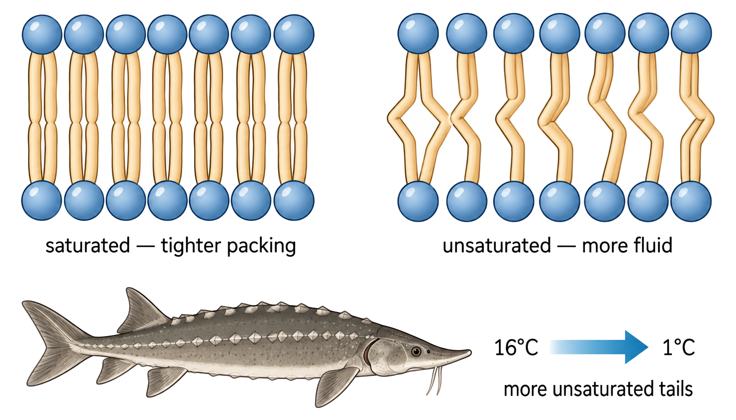
Cis double bonds create bends in fatty-acid tails, reducing packing and generally increasing membrane fluidity at a given temperature.

Bent tails make neighboring phospholipids fit less tightly, so less thermal energy is needed for them to move past each other. Chain length and the number of double bonds also affect the result.

Compare two membranes at the same temperature: the one with more cis unsaturation usually has looser packing and greater fluidity.

A membrane rich in bent tails can remain flexible in cool conditions where a membrane of straight saturated tails would become more rigid.

Do not treat unsaturation as the only control: temperature, chain length and cholesterol can modify the prediction.

Fatty acid composition and fluidity

HL only

Assessment in practice

1 marks
How it is assessed

This objective is assessed through structured response, commonly using Identify / Outline.

Command terms

Identify / Outline

What earns marks

Build the answer around this relationship: Unsaturated fatty acid tails increase membrane fluidity by reducing tight packing.

Watch for

Reversing the effects of saturated and unsaturated fatty acids on membrane fluidity.

Representative question

Question 1

[Maximum number: 1]

Outline the effect of fatty acids on the fluidity of membranes.

Cholesterol Buffers Membrane Fluidity

HL only

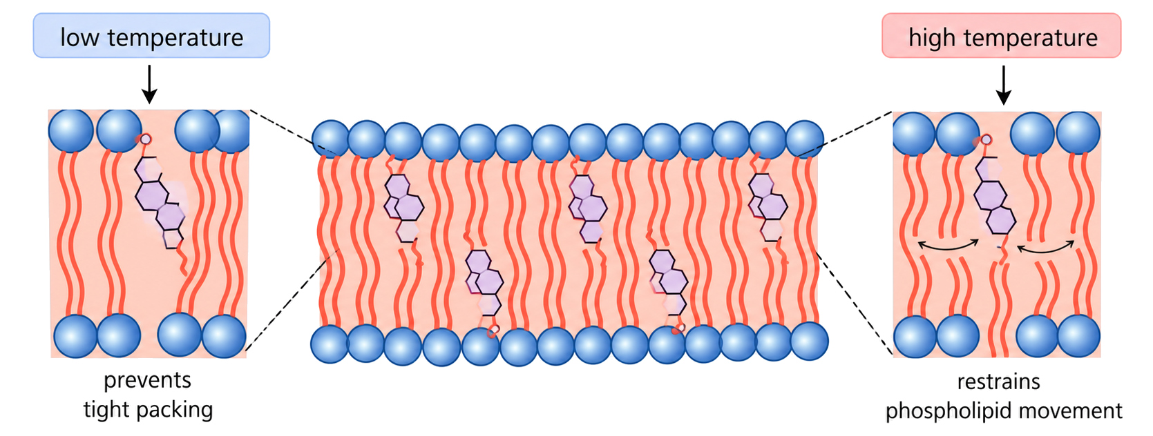
Cholesterol fits between phospholipids and helps prevent membranes becoming too rigid when cold or too fluid when warm.

Its small polar hydroxyl group sits near phospholipid heads while its rigid hydrophobic body restricts tail movement. This makes cholesterol a buffer rather than a simple ‘fluidity increaser’.

State the condition first: cold membranes need prevention of tight packing; warm membranes need restraint of excessive movement.

Adding cholesterol can stop a warm membrane becoming excessively leaky, but in cold conditions it can prevent phospholipids packing into a solid.

Saying cholesterol always increases fluidity is incomplete; its effect depends on temperature and the surrounding lipid composition.

Cholesterol and fluidity

HL only

Assessment in practice

1 marks
How it is assessed

This objective is assessed through structured response, commonly using Identify / State / Outline.

Command terms

Identify / State / Outline

What earns marks

The repeated one-mark rows make the same central demand from several angles: identify cholesterol by its membrane role.

Watch for

Giving only a vague stabilizing role without mentioning fluidity or permeability.

Representative question

Question 1

[Maximum number: 1]

Outline the effect of cholesterol on the fluidity of membranes.

Fluid Membranes Can Bend into Vesicles

HL only

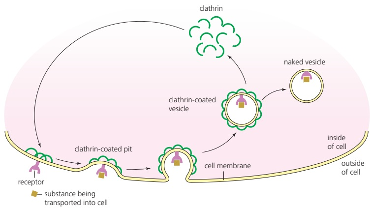
Membrane fluidity lets a bilayer curve, fuse and pinch off, allowing vesicles to form and move material between compartments.

Lipids can move laterally and the two leaflets can rearrange their shape. Proteins and cytoskeletal forces help control budding and fusion, while the bilayer’s continuity preserves a sealed aqueous compartment.

Follow the event: membrane bends → a bud forms → neck closes or fuses → cargo remains enclosed by bilayer.

A secretory vesicle can bud from an internal membrane and later fuse with the cell surface, releasing cargo outside.

Vesicle formation is not simple leakage. A sealed bilayer surrounds the cargo; uncontrolled holes would not preserve compartmentalization.

Membrane fluidity and vesicles

HL only

Assessment in practice

1–5 marks
How it is assessed

This objective is assessed through structured response, commonly using Describe / Identify / State.

Command terms

Describe / Identify / State / Explain

What earns marks

Build the answer around this relationship: Fluid membranes can change shape to form vesicles.

Watch for

Naming endocytosis without describing membrane invagination and vesicle formation.

Representative question

Question 1

[Maximum number: 5]

Explain how vesicles are used by cells to move materials.

Gated Ion Channels Open Only Under a Signal

HL only

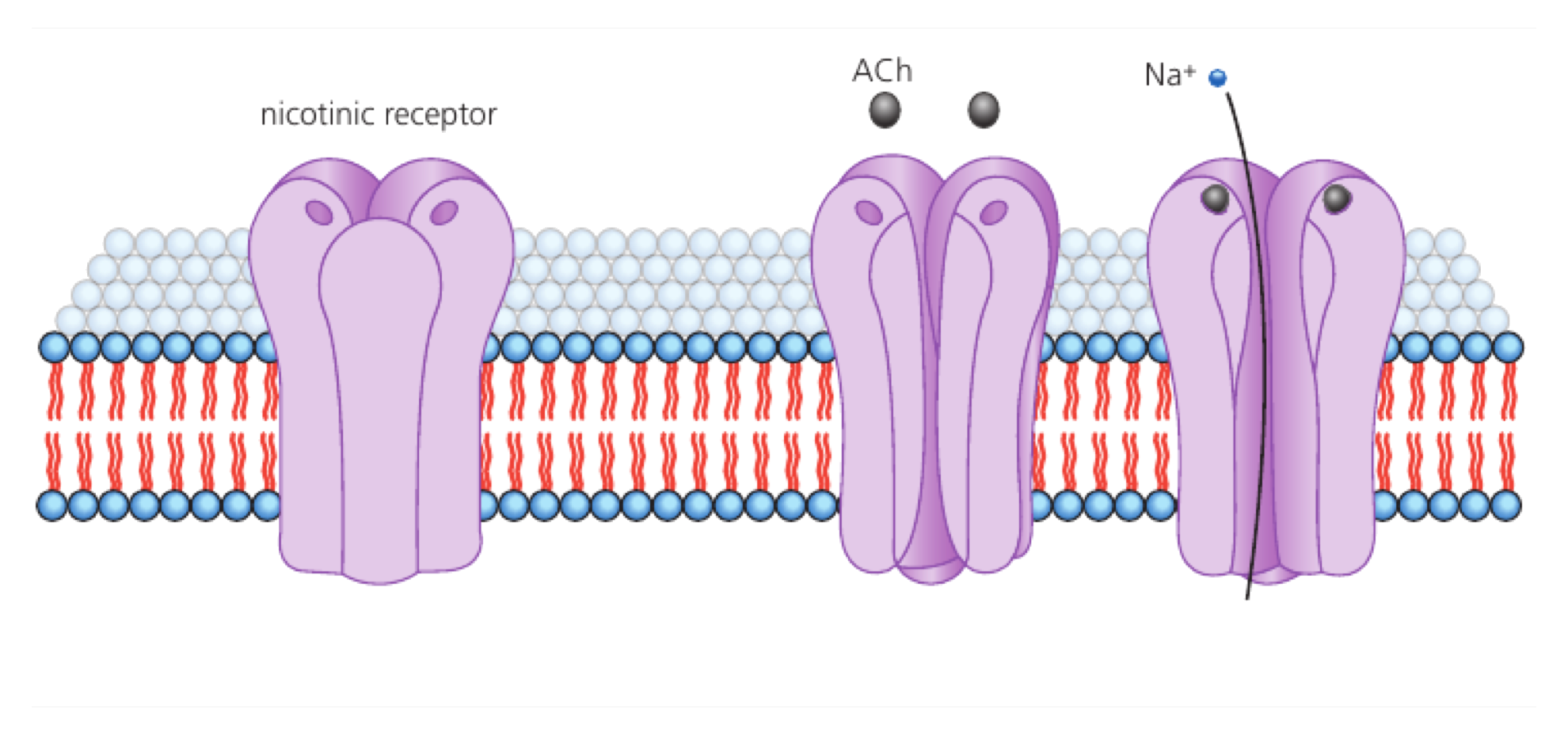
Gated ion channels change between closed and open states when a specific stimulus changes their conformation, allowing selected ions to cross down their electrochemical gradient.

The gate links stimulus detection to pore opening. Ion selectivity comes from the pore’s chemistry, while the electrochemical gradient determines the direction of movement after opening.

Separate three questions: what opens the gate, which ion fits, and which way the gradient drives it.

A voltage-gated sodium channel opens after membrane potential changes, then sodium moves according to its electrochemical gradient.

Opening a channel does not itself pump ions uphill or determine a universal direction; gradient and selectivity still matter.

Gated ion channels in neurons

HL only

Assessment in practice

1–3 marks
How it is assessed

This objective is assessed through structured response, commonly using Explain / State / Identify.

Command terms

Explain / State / Identify

What earns marks

Build the answer around this relationship: Voltage-gated potassium channels open in response to membrane-potential changes.

Watch for

Swapping neurotransmitter-gated acetylcholine receptors with voltage-gated potassium channels.

Representative question

Question 1

[Maximum number: 3]

Explain the action of the voltage-gated potassium channel during a nerve impulse.

The Sodium–Potassium Pump Builds an Ion Gradient

HL only

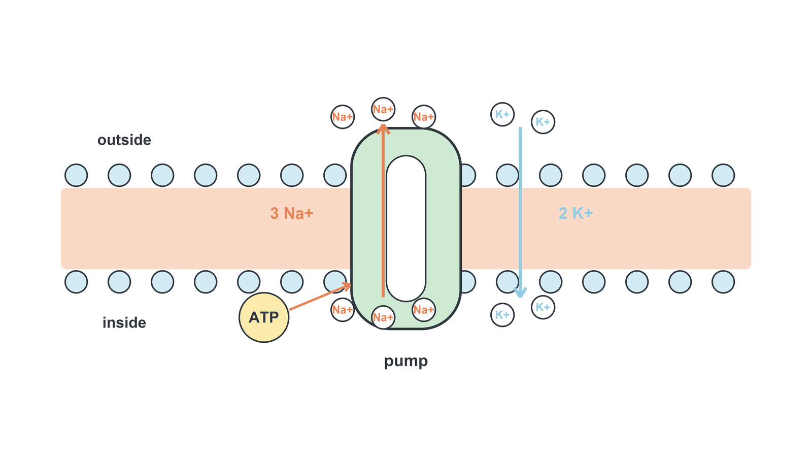
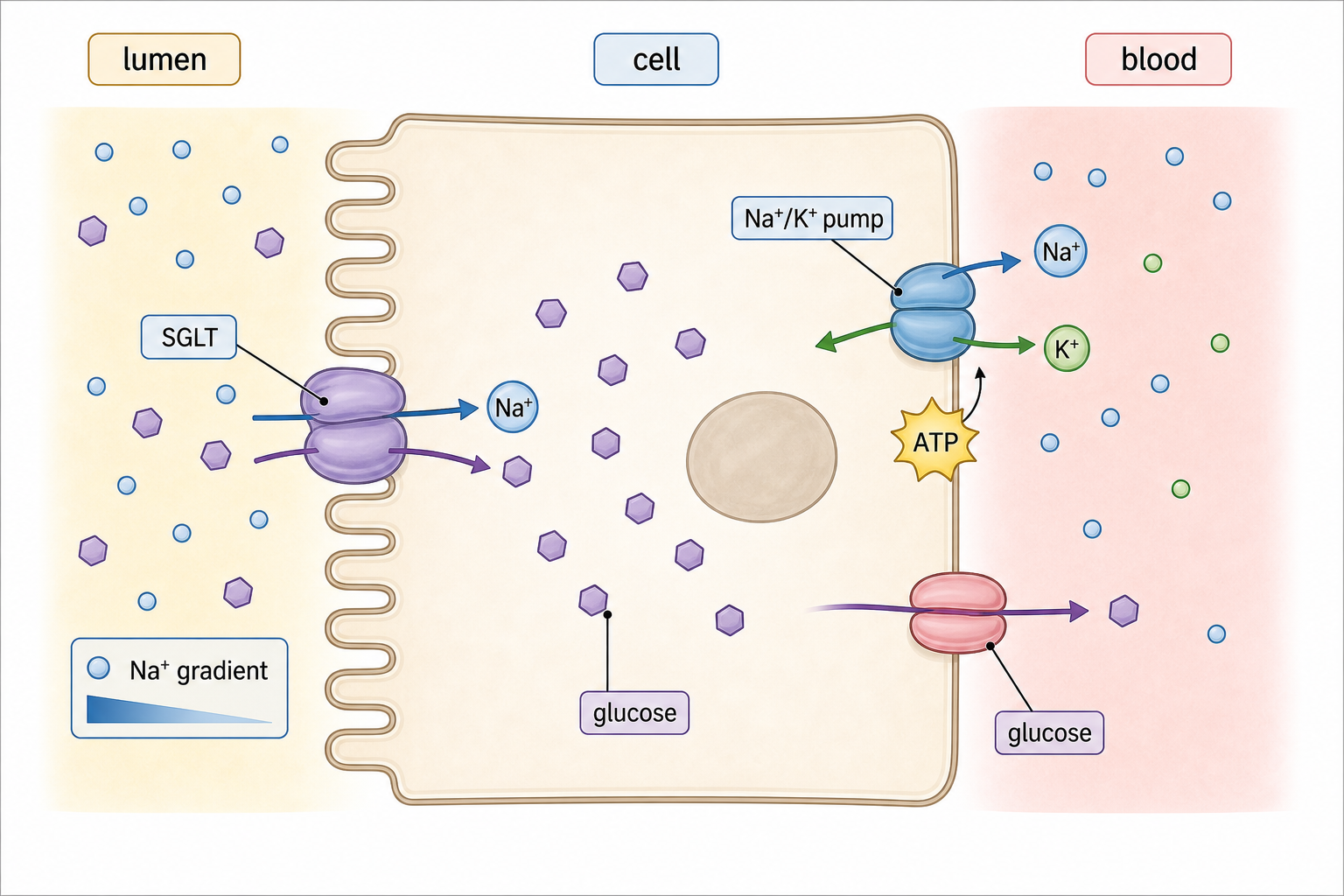
The sodium–potassium pump uses ATP to move sodium out of a cell and potassium into it against their gradients, typically exchanging three sodium ions for two potassium ions per cycle.

ATP phosphorylation changes the pump’s shape so binding sites face alternate sides. Repeated cycles maintain unequal ion distributions that can later drive electrical signals or cotransport.

Track one cycle: three Na⁺ bind inside → ATP-powered shape change releases them outside → two K⁺ bind outside → reset releases K⁺ inside.

If the pump stops, diffusion gradually reduces the sodium and potassium gradients even though channels may still function.

The pump creates gradients; it is not the same as a channel that lets ions flow down those gradients.

Sodium-potassium pumps

HL only

Assessment in practice

1 marks
How it is assessed

This objective is assessed through structured response, commonly using Identify / Suggest / Analyse.

Command terms

Identify / Suggest / Analyse

What earns marks

Build the answer around this relationship: Sodium-potassium pumps use ATP for active transport.

Watch for

Reversing sodium and potassium movement across the membrane.

Representative question

Question 1

[Maximum number: 2]

Analyse the graph to obtain two conclusions about the concentration of sodium-potassium pumps.
1.
2.

Sodium–Glucose Cotransport Uses One Gradient to Move Another Solute

HL only

A sodium–glucose cotransporter couples downhill sodium movement to uphill glucose movement across a membrane.

The transporter binds both solutes and changes shape only when the correct combination is present. The sodium gradient, maintained by the sodium–potassium pump, supplies the energy indirectly.

Trace the coupling: sodium moves down its gradient, glucose is carried against its gradient, and the pump restores sodium conditions. Both solutes must bind for the cycle to proceed.

In an intestinal cell, sodium entry can bring glucose in even when glucose concentration is already higher inside.

This is secondary active transport: the cotransporter does not split ATP directly, but it depends on an ATP-maintained sodium gradient.

Sodium-glucose cotransporters

HL only

Assessment in practice

1 marks
How it is assessed

This objective is assessed through multiple choice, commonly using Identify.

Command terms

Identify

What earns marks

Teaching content should connect that answer to the mechanism: sodium moves down its gradient, glucose is carried with it, and the cell later moves glucose onward.

Representative question

Question 1

[Maximum number: 1]

What transport method is used in the reabsorption of glucose in the proximal convoluted tubule of the kidney?

A

Diffusion

B

Osmosis

C

Endocytosis

D

Active transport

Cell Adhesion Molecules Link Neighboring Cells

HL only

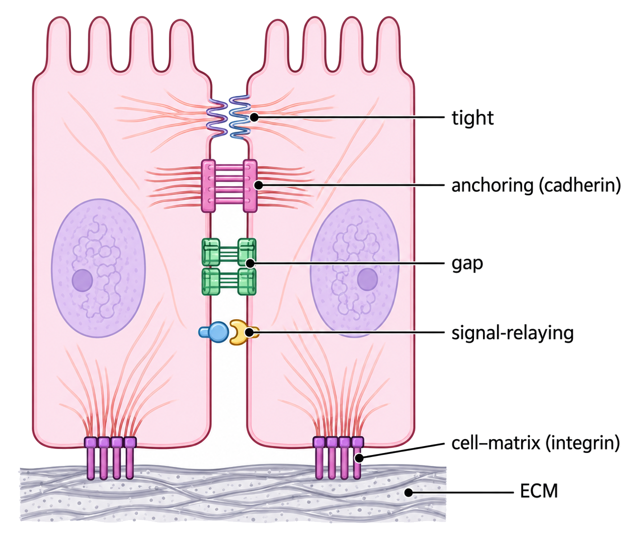
Cell adhesion molecules are membrane proteins that bind matching molecules on neighboring cells or the extracellular matrix, helping organize tissues.

Specific extracellular domains recognize partners, while intracellular regions connect to cytoskeletal or signaling systems. Adhesion therefore provides both physical attachment and information about tissue structure.

Explain an adhesion effect by naming: binding partner, extracellular contact, and intracellular consequence.

If CAM binding is weakened, epithelial cells may separate more easily because the molecular link to neighboring cells and the cytoskeleton is reduced.

Adhesion is not permanent glue. CAM binding can be regulated, and different CAMs have different partners and tissue roles.

Cell adhesion molecules (CAMs)

HL only

Assessment in practice

1 marks
How it is assessed

This objective is assessed through multiple choice, commonly using Identify.

Command terms

Identify

What earns marks

Build the answer around this relationship: Cell adhesion molecules help cells attach to other cells or extracellular matrix.

Representative question

Question 1

[Maximum number: 1]

Animal cells often secrete glycoproteins as extracellular components. What is a role of these glycoproteins?

A

Adhesion

B

Additional energy reserve

C

Membrane fluidity

D

Water uptake

Fluidity, Neurons, Cotransport, Adhesion

HL only

The HL extension asks how membrane structure becomes dynamic cell behaviour. Fatty acid saturation and cholesterol tune fluidity. Fluid membranes form and fuse vesicles. Gated ion channels and sodium-potassium pumps create nerve-cell gradients and electrical responses. Sodium-dependent glucose cotransport uses a sodium gradient to move glucose indirectly against its gradient. Adhesion molecules organize tissues.

  • Unsaturated tails increase fluidity; saturated tails pack closely.
  • Cholesterol buffers animal membrane fluidity at low and high temperature.
  • Fluid membranes allow endocytosis and exocytosis.
  • Gated channels and sodium-potassium pumps support nerve-cell membrane potentials.
  • Sodium-glucose cotransport is indirect active transport.
  • Cadherins, integrins, and junctions organize tissues.
ConceptIB Biology HL