Question 1
[Maximum number: 5]
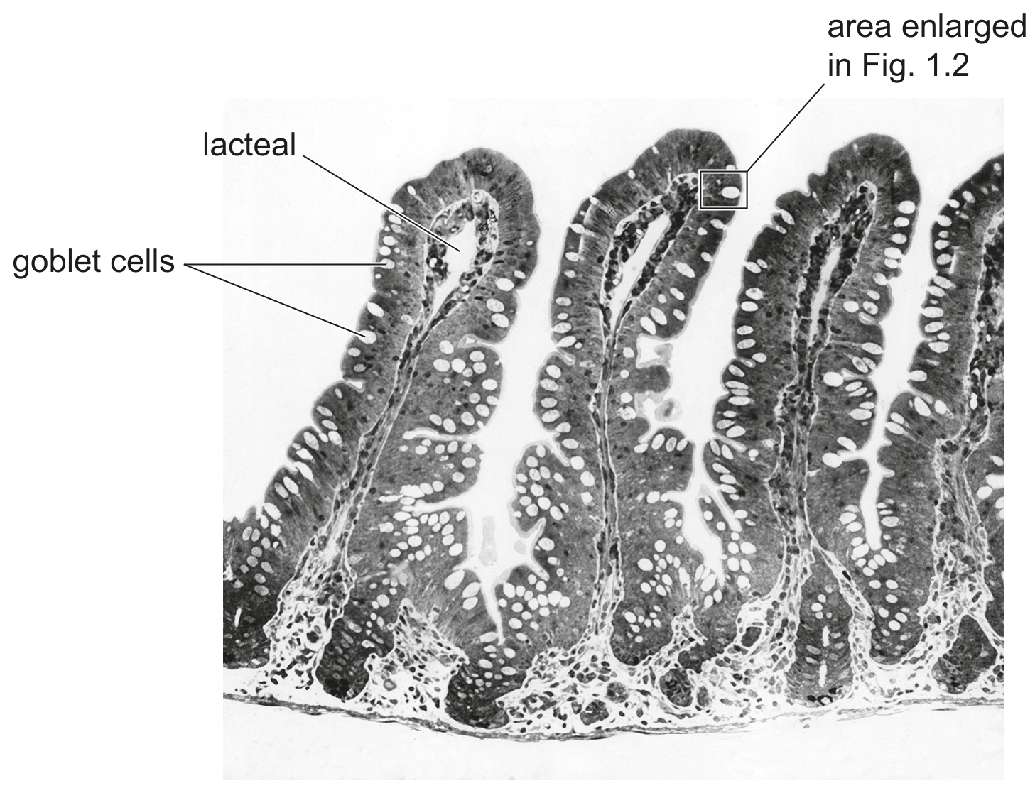
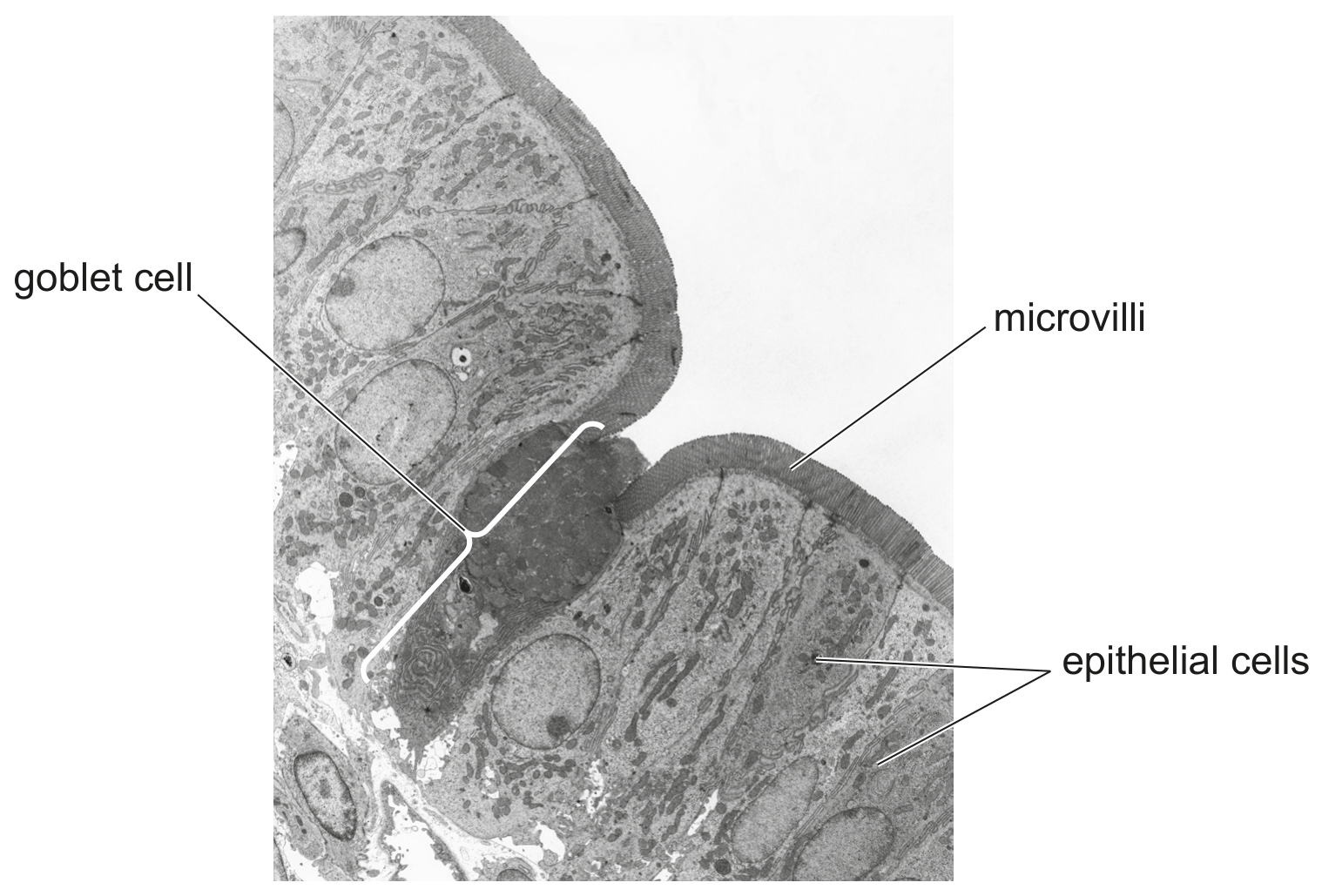
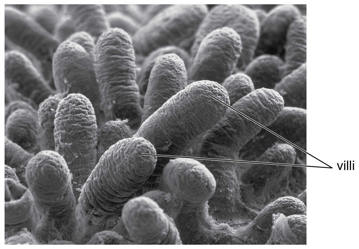
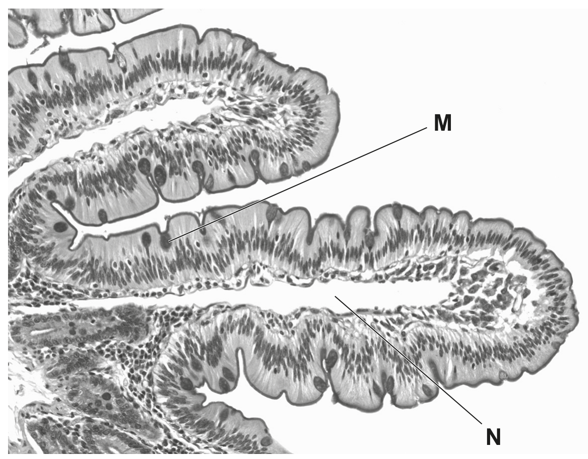
Fig. 1.1 and Fig. 1.2 show two images of villi.
Fig. 1.1 shows a surface view of many villi viewed through a scanning electron microscope.
Fig. 1.2 shows a section of one villus viewed through a light microscope.
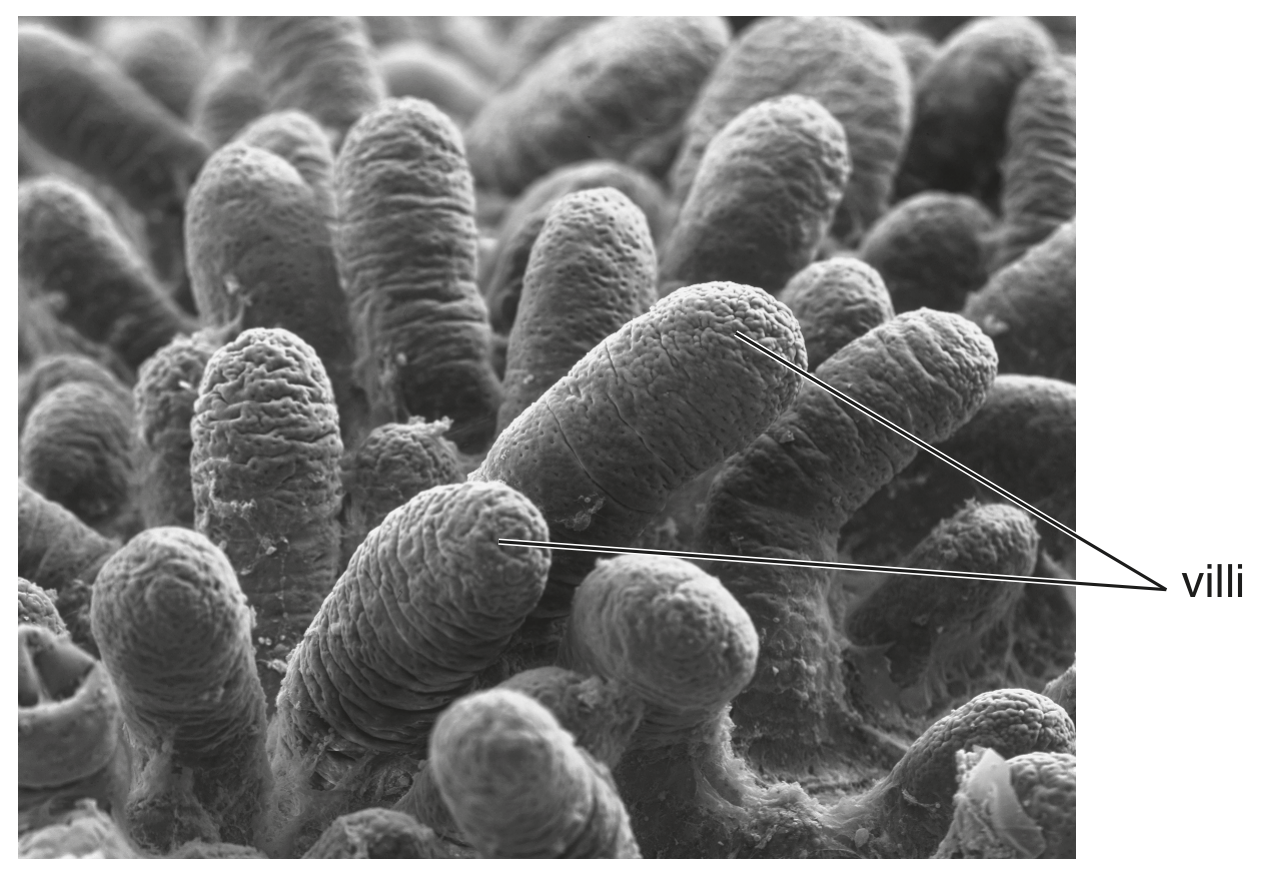
Fig. 1.1
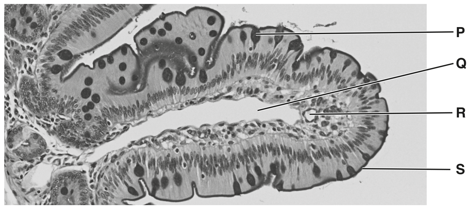
Fig. 1.2
Villi are found in the small intestine.
Question 1(a)
(a)
State the function of villi.
[ 1 ]
Question 1(b)
(b)
Identify and describe two of the labelled components of a villus.
Use the letters in Fig. 1.2 in your answer.
[ 4 ]