EduNinja

IGCSE Biology11.1 Gas exchange in humansQuestion Bank

Question 1

Question 1(a)

(a)

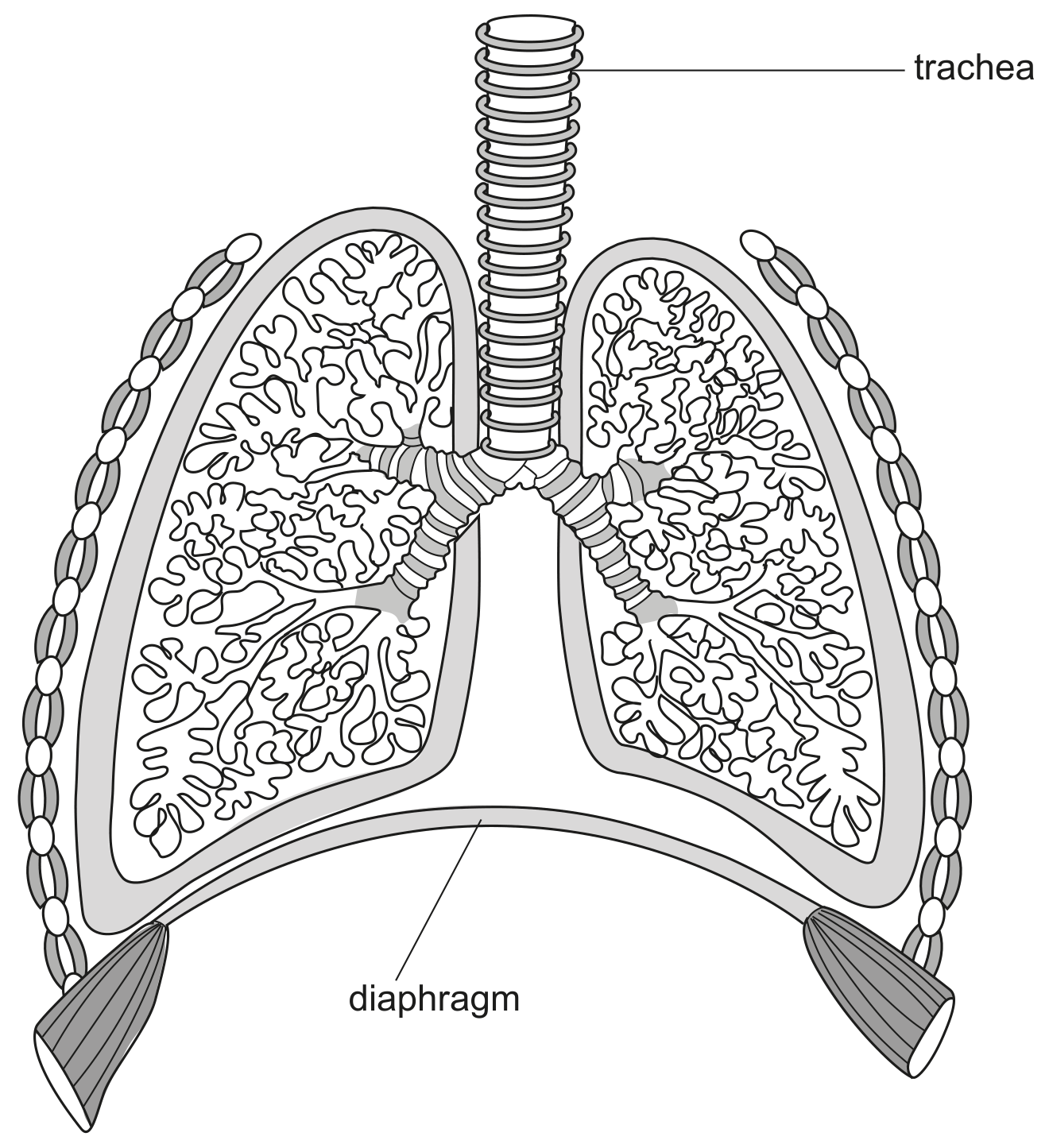
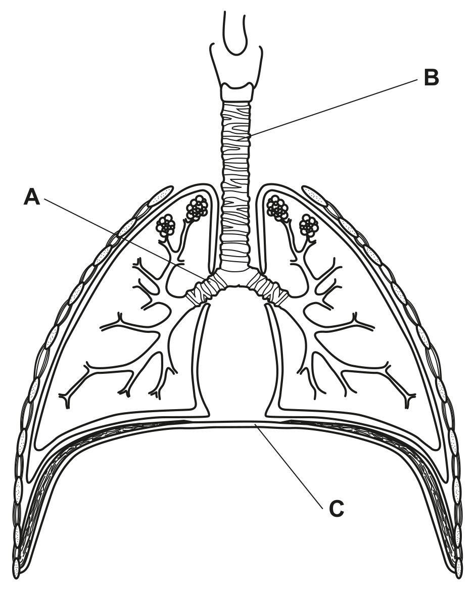
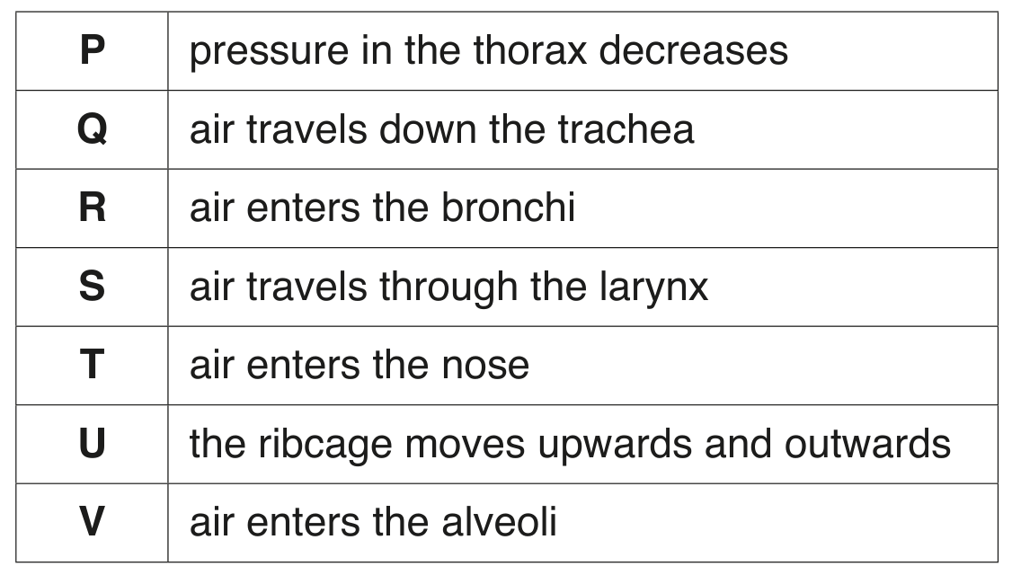
Fig. 1.1 shows the human gas exchange system. The functions of the parts of the gas exchange system are given in Table 1.1.
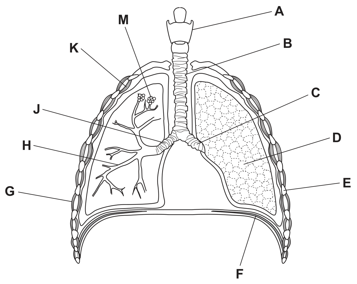
Fig. 1.1

Fig. 1.1

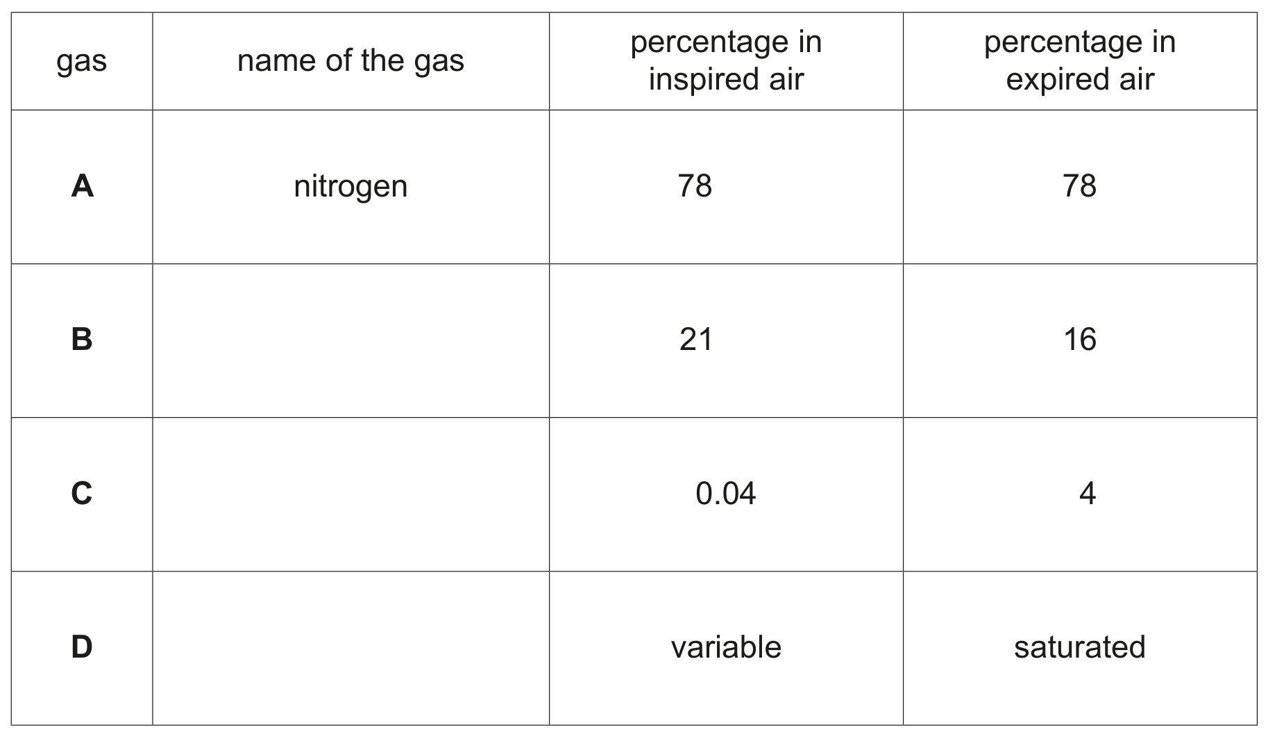
Complete Table 1.1. One row has been done for you.
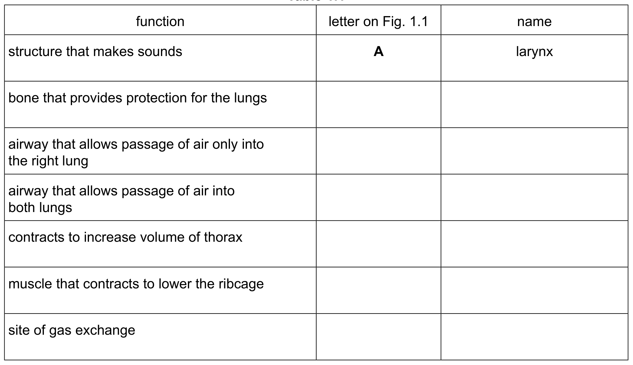
Table 1.1

Table 1.1

[ 6 ]

Question 1(b)

(b)

The gas exchange system contains cartilage.

Describe the function of cartilage in the gas exchange system.

[ 2 ]

Question 1(c)

(c)

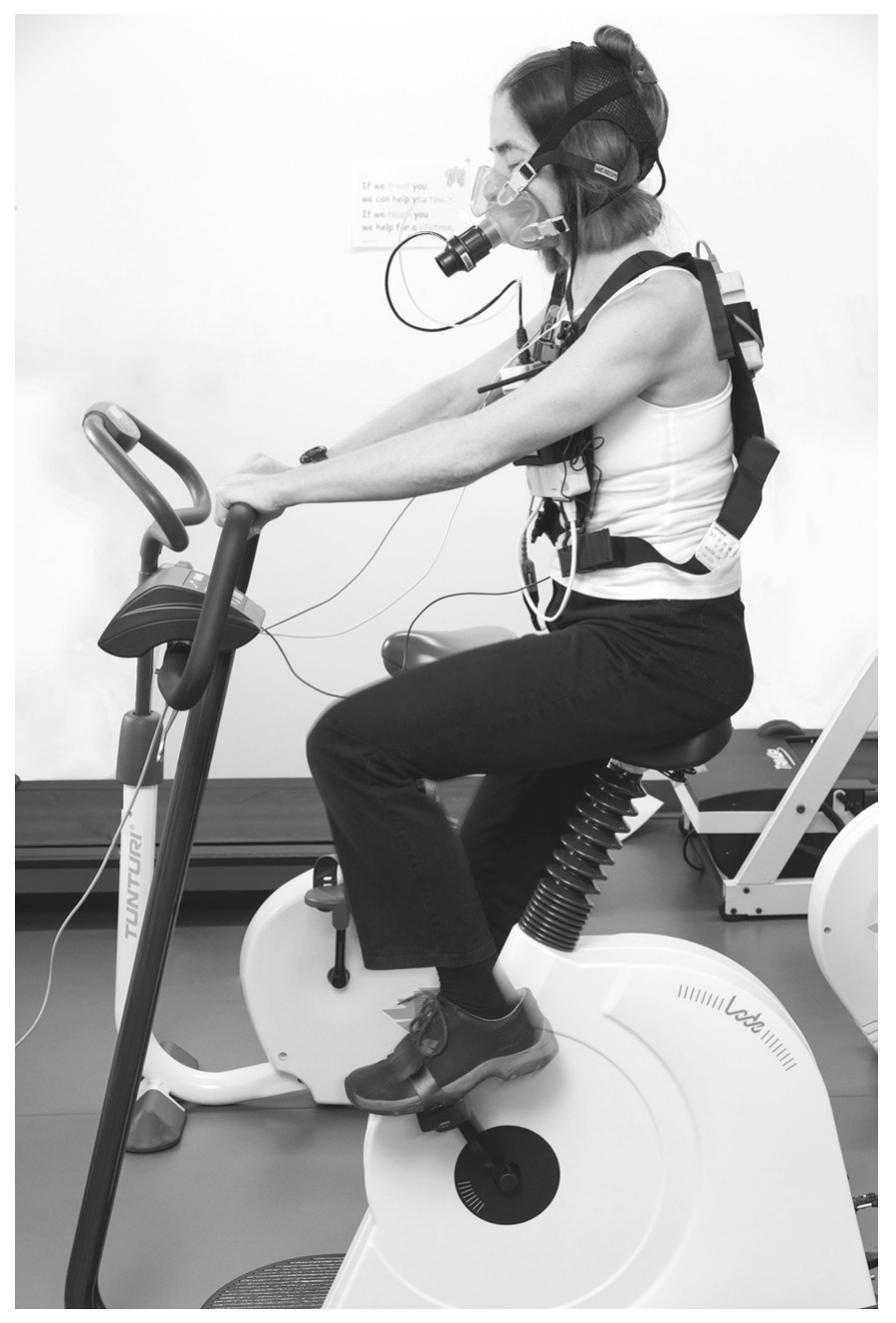
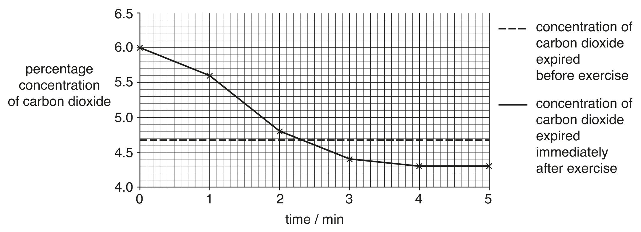
Soon after starting physical activity the concentration of carbon dioxide in the blood increases.

[ 4 ]

Question 1(c)(ii)

(i)

State the effect on breathing of an increase in carbon dioxide concentration in the blood.

[ 1 ]

Question 1(c)(iii)

(ii)

Explain how this effect on breathing is coordinated.

[ 3 ]

Question 1

Question 1(a)

Question 1(a)(i)

(a)
(i)

State the name of the gas exchange surface in humans.

[ 1 ]

Question 1(a)(ii)

(ii)

State two features of the gas exchange surface in humans.

1
2

[ 2 ]

Question 1(b)

(b)

Fig. 1.1 is a diagram of the gas exchange system in humans.

Fig. 1.1

Fig. 1.1

[ 5 ]

Question 1(b)(i)

(i)

Draw a label line and the letter X on Fig. 1.1 to identify an external intercostal muscle.

[ 1 ]

Question 1(b)(ii)

(ii)

State the name of the tissue that forms C-shaped structures in the wall of the trachea and state its function.
name
function

[ 2 ]

Question 1(b)(iii)

(iii)

Describe the effects on the thorax of contraction of the diaphragm.

[ 2 ]

Question 1(c)

(c)

Table 1.1 compares the composition of inspired and expired air.

Table 1.1

Table 1.1

[ 6 ]

Question 1(c)(i)

(i)

Complete Table 1.1 by writing the names of gases B, C and D.

[ 3 ]

Question 1(c)(ii)

(ii)

For gas B and gas C, explain the differences in the percentages shown in Table 1.1 between inspired and expired air.

[ 3 ]

Question 1

[Maximum number: 3]

Fig. 1.1 shows several villi from the ileum, which is part of the small intestine.

Question 1(c)

(a)

Goblet cells provide protection for the epithelial cells that line the intestine.

[ 3 ]

Question 1(c)(i)

(i)

State the name of the protective substance produced by goblet cells.

[ 1 ]

Question 1(c)(ii)

(ii)

Suggest why a protective substance is necessary in the intestines.

[ 2 ]

Question 1

Question 1(b)

(a)

Fig. 1.1 is a micrograph of part of some fish gills.

Fig. 1.1

Fig. 1.1

Fish gills are adapted for gas exchange by diffusion.

[ 1 ]

Question 1(b)(ii)

(i)

Suggest one adaptation, visible in Fig. 1.1, that shows that fish gills are efficient structures for gas exchange by diffusion.

[ 1 ]

Question 1

[Maximum number: 8]

The gas exchange system is one of the organ systems of the human body.

Fig. 1.1 shows parts of the gas exchange system during breathing in and breathing out.

Fig. 1.1 shows parts of the gas exchange system during breathing in and breathing out.

Question 1(a)

(a)

Complete Table 1.1 to show:
- the functions of the diaphragm and the intercostal muscles during breathing in and breathing out
- the pressure changes in the thorax.

Use these words:
```
contract
relax
increases
decreases.
```

Table 1.1

Table 1.1

[ 4 ]

Question 1(b)

(b)

Fig. 1.2 shows part of the gas exchange surface of a human.

Fig. 1.2

Fig. 1.2

State two features of the gas exchange surface that are visible in Fig. 1.2.

1

2

[ 2 ]

Question 1(c)

(c)

The cells labelled X on Fig. 1.2 form a tissue.

[ 2 ]

Question 1(c)(ii)

(i)

Cartilage is another tissue found in the gas exchange system.

State the functions of cartilage in the gas exchange system.

[ 2 ]

Question 1

Question 1(a)

(a)

Fig. 1.1 is a diagram of the human gas exchange system.

Fig. 1.1

Fig. 1.1

[ 7 ]

Question 1(a)(i)

(i)

Identify the structures labelled A, B and C in Fig. 1.1.

A

B
C

[ 3 ]

Question 1(a)(ii)

(ii)

Explain how the structures in the gas exchange system cause inspiration.

[ 4 ]

Question 1

[Maximum number: 9]

Fig. 1.1 shows two photomicrographs of a cross-section of trachea.

Fig. 1.1

Fig. 1.1

Question 1(a)

(a)

The open space at the centre of the trachea is labelled A in Fig. 1.1.

Air travels into and out of the trachea through this open space.
State a gas that is at a higher concentration in expired air than in inspired air.

[ 1 ]

Question 1(b)

(b)

Describe and explain the functions of the structures or substances labelled B to E in Fig. 1.1. Use the letters in Fig. 1.1 in your answer.

[ 4 ]

Question 1(c)

(c)

Fig. 1.2 shows some events during inspiration.

Fig. 1.2

Fig. 1.2

[ 4 ]

Question 1(c)(i)

(i)

Put the events shown in Fig. 1.2 into the correct sequence. Two have been done for you.

Table
[ 2 ]

Question 1(c)(ii)

(ii)

Suggest why alveoli have thin walls.

[ 2 ]

Question 2

[Maximum number: 2]

The concentration of atmospheric carbon dioxide has increased considerably in recent years.

Question 2(d)

(a)

When the scientists were working in the plot with a carbon dioxide concentration of 550 ppm , their breathing rates were higher than when they worked in the other plot.

Suggest why their breathing rates were higher.

[ 2 ]

Question 2

[Maximum number: 10]

A group of students planned an investigation to determine the effects of physical activity on breathing rate.

Question 2(a)

(a)

Describe how the students could measure their breathing rates.

[ 2 ]

Question 2(b)

(b)

The students measured their breathing rates before physical activity and every minute for five minutes after cycling around the school field.

Write a hypothesis for their investigation.

[ 2 ]

Question 2(c)

(c)

Fig. 2.1 shows a woman on a stationary bicycle. The mask fitted over her nose and mouth measures the composition of the air she breathes out.

Fig. 2.1

Fig. 2.1

Fig. 2.2 shows the concentration of carbon dioxide in the air expired by the woman in the five minutes after she stopped exercising.

The dashed line on the graph shows the concentration of carbon dioxide in her expired air when she was at rest, before she began to exercise.

Fig. 2.2

Fig. 2.2

Describe and explain the results of the investigation shown in Fig. 2.2.
Use the data in Fig. 2.2 in your answer.

[ 6 ]

Question 3

Question 3(a)

Question 3(a)(i)

(a)
(i)

Complete Table 3.1 by writing in the percentages of carbon dioxide and oxygen in inspired air and in expired air.

Table 3.1

Table 3.1

[ 2 ]

Question 3(a)(ii)

(ii)

A scientist measured the number of dust particles in inspired air and in expired air.

They found fewer dust particles in expired air.
Suggest a reason for their observation.

[ 1 ]

Question 3(b)

(b)

Fig. 3.1 is a diagram of alveoli and associated blood vessels.

Fig. 3.1

Fig. 3.1

[ 2 ]

Question 3(b)(iv)

(i)

State the location and function of cartilage in the breathing system.
location
function

[ 2 ]

Question 3(c)

(c)

A student measured the rate and depth of breathing of an athlete for 30 seconds at rest. The data are shown in Fig. 3.2.

Fig. 3.2

Fig. 3.2

[ 6 ]

Question 3(c)(i)

(i)

Using the information in Fig. 3.2, calculate the rate of breathing at rest. breaths per minute

The measurements were repeated while the athlete was running on a treadmill.
The data are shown in Fig. 3.3.

Fig. 3.3

Fig. 3.3

[ 1 ]

Question 3(c)(ii)

(ii)

Using the information in Fig. 3.3, calculate the volume of air inspired in one breath from 25 seconds. dm3\mathrm{dm}^{3}

[ 1 ]

Question 3(c)(iii)

(iii)

Explain the effect of exercise on the rate and depth of breathing shown in Fig. 3.2 and Fig. 3.3.

[ 4 ]
0 selected