Question 2
[Maximum number: 1]
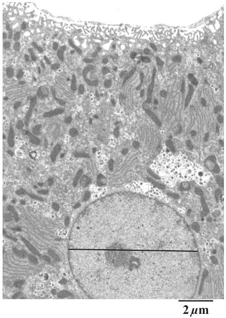
The electron micrograph shows part of a cell. Which organelle is the site of aerobic respiration?
 EduNinja
The electron micrograph shows part of a cell. Which organelle is the site of aerobic respiration?
The image shows an electron micrograph of a cell.
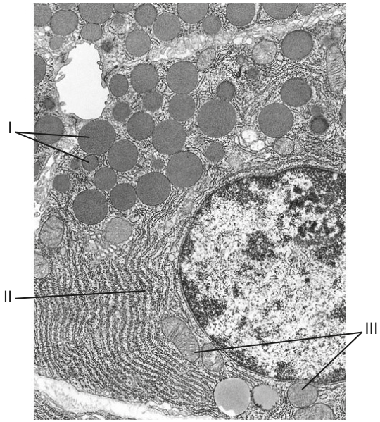
Which organelles correspond to the labels in the electron micrograph of this cell?
1
II
III
secretory vesicles
Golgi apparatus
mitochondria
mitochondria
Golgi apparatus
secretory vesicles
secretory vesicles
rough endoplasmic reticulum
mitochondria
mitochondria
rough endoplasmic reticulum
secretory vesicles
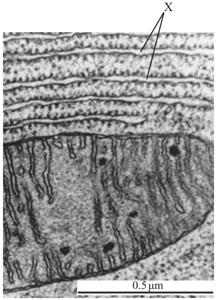
What is the function of the organelles labelled X ?
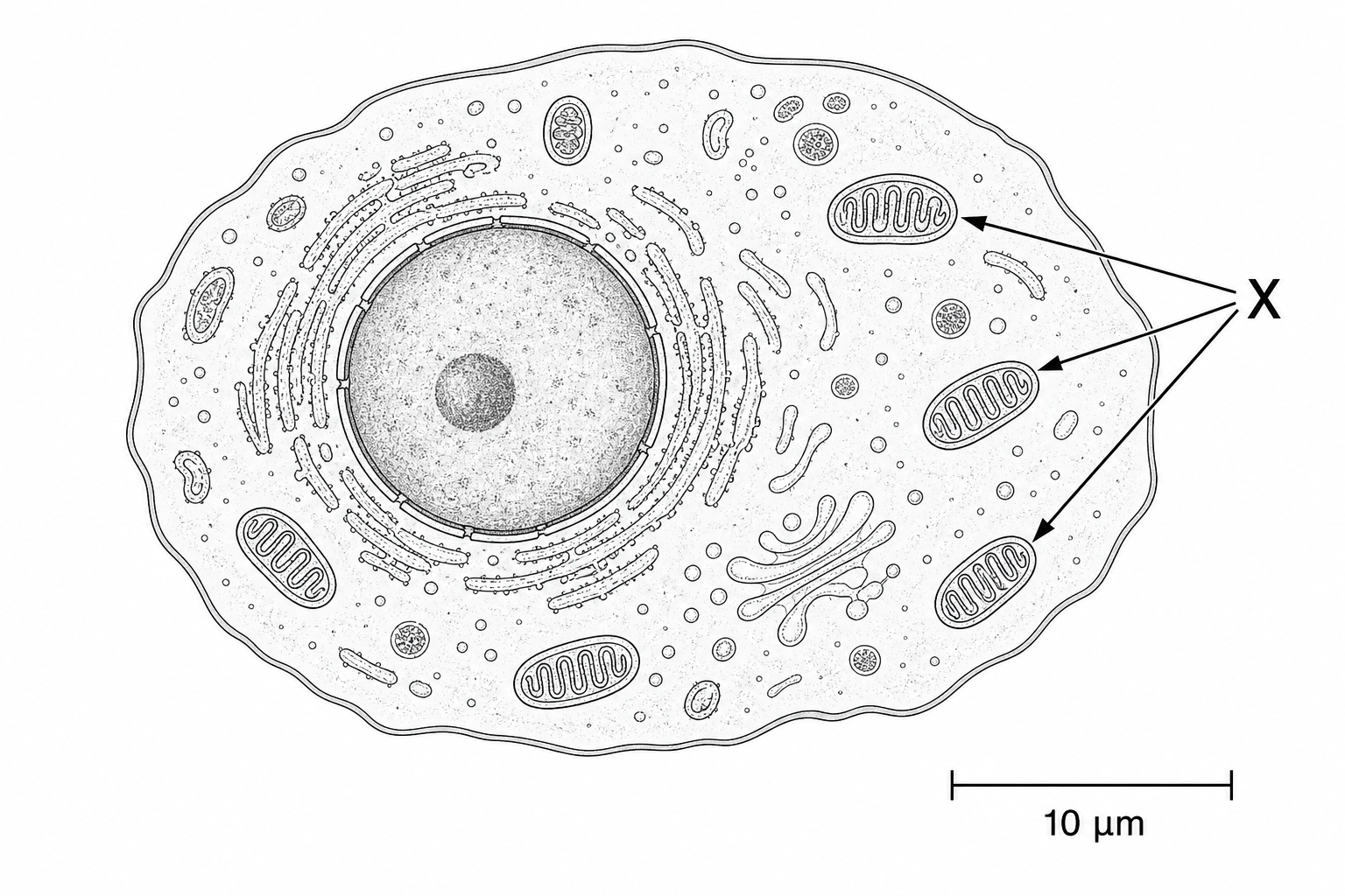
Glycolysis
Polypeptide formation
Aerobic cell respiration
Protein transport
The diagram shows how vesicles are used to transport materials in a cell.
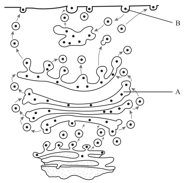
State the name of organelle A .
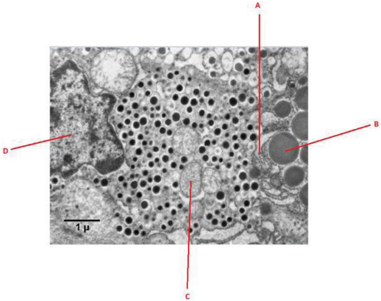
The electron micrograph shows part of a eukaryotic cell.
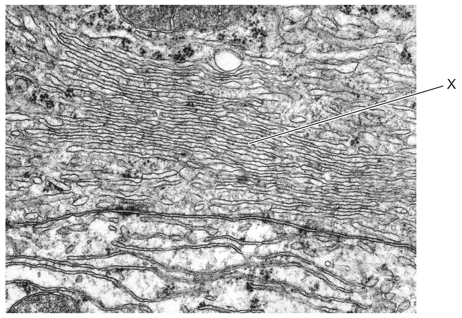
What is the organelle labelled X ?
Mitochondrion
Smooth endoplasmic reticulum
Lysosome
Nucleus
The image is a transmission electron micrograph of part of two adjacent pancreatic cells.
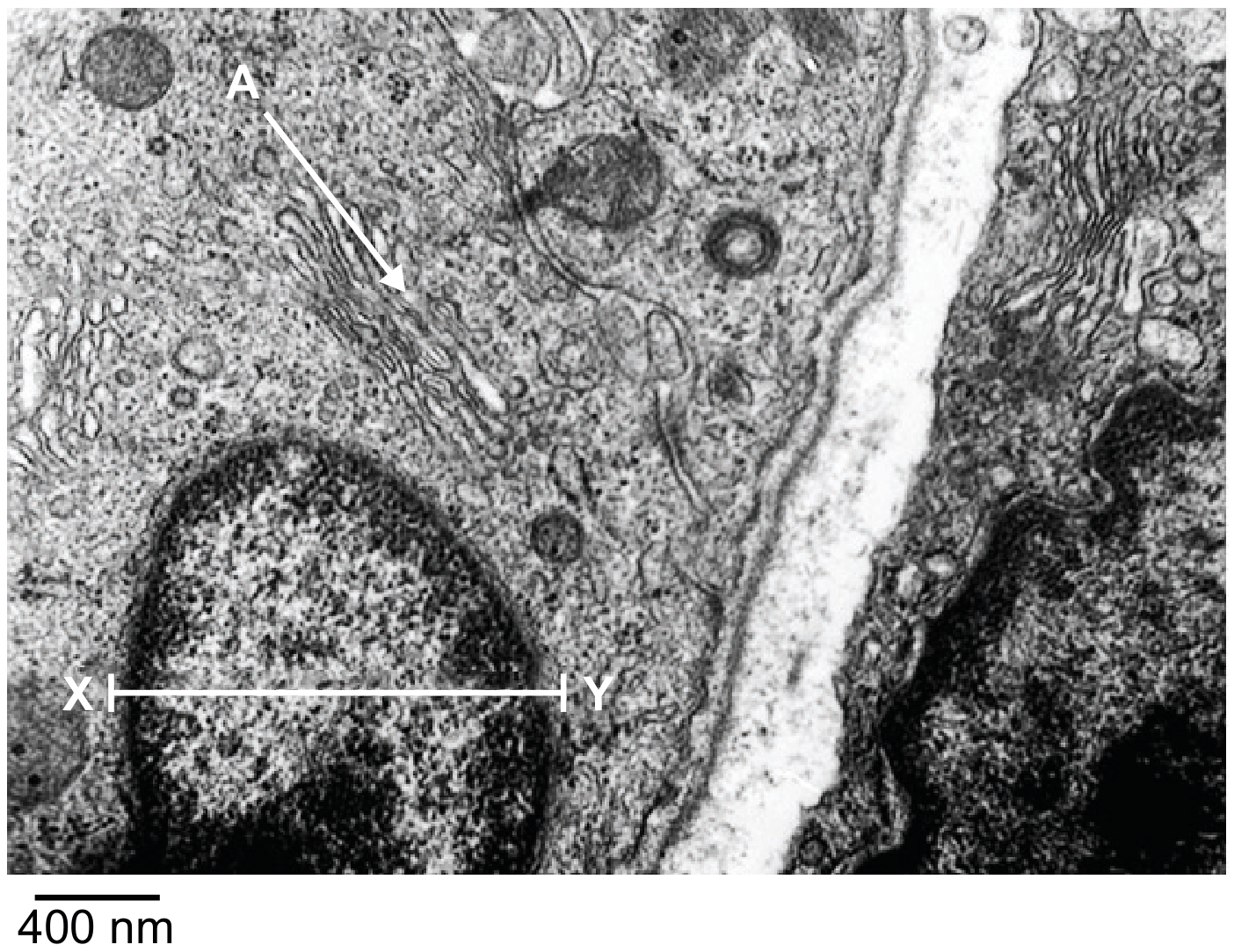
Identify the structure labelled A.
Which of the following is considered a cell organelle?
Cell wall
Lysosome
Cytoskeleton
Cytoplasm
Identify which electron micrograph shows a mitochondrion, providing one observation to support your choice.
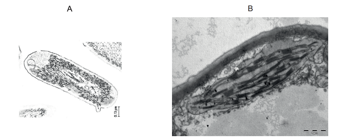
C
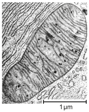
The following shows an electron micrograph of a liver cell.
The electron micrograph is a higher magnification of a liver cell.
State its main function.
The diagram shows the structure of part of a fungus.
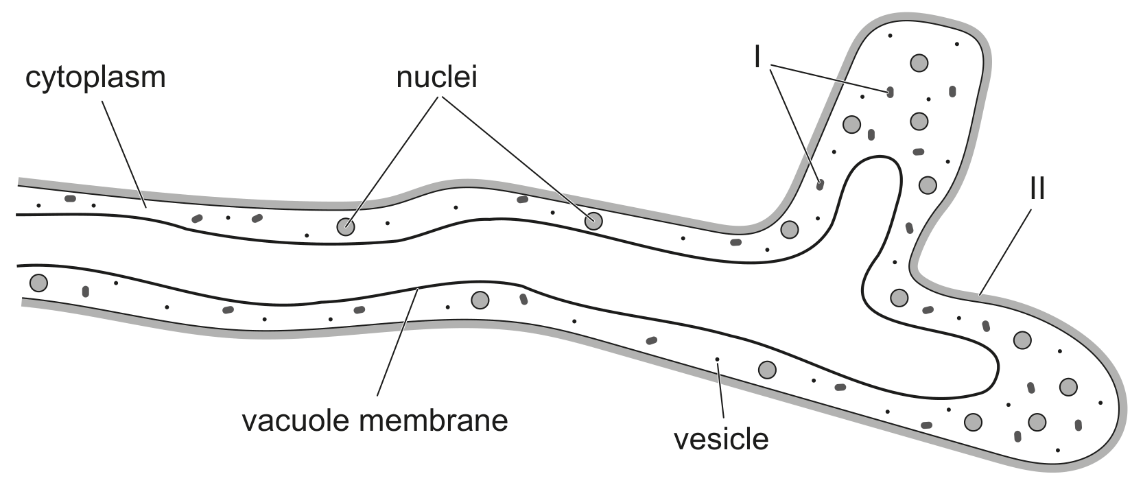
The structures labelled I are membrane-bound organelles. Deduce, giving a reason, what these organelles could be.