Question 1
[Maximum number: 2]
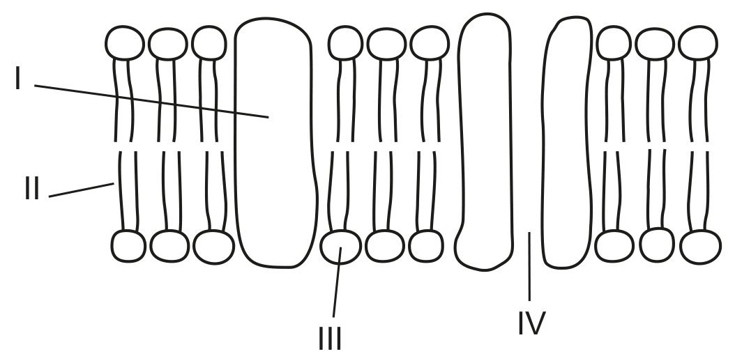
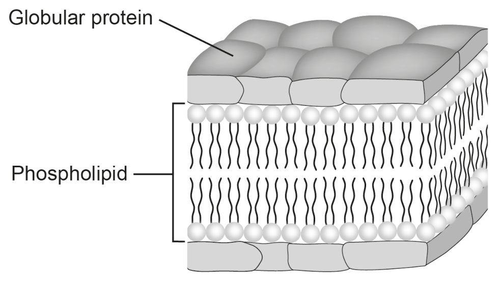
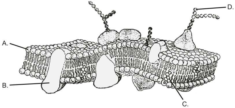
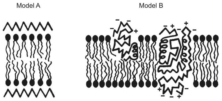
Two models of plasma membrane structure are shown.
Question 1(b)
Question 1(b)(i)
(a)
(i)
Label the model A diagram to show a region of protein.
[ 1 ]
Question 1(b)(ii)
(ii)
Label the model B diagram to show a phospholipid.
[ 1 ]