EduNinja

IB Biology SLA2.2 Cell structureQuestion Bank

Question 1

[Maximum number: 1]

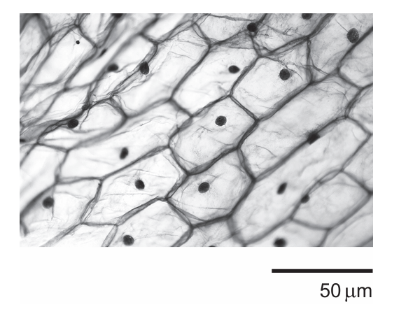
The image was obtained with a mobile phone camera pointing down the eyepiece tube of a light microscope. It shows onion (Allium cepa) epidermal cells after immersion in a hypertonic salt solution during an experiment on osmosis.
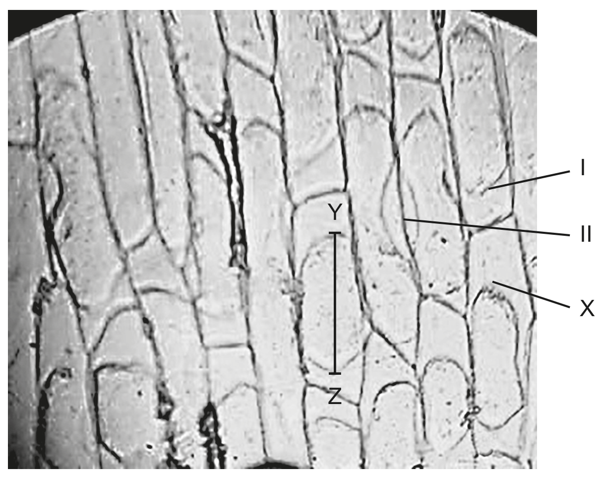
\(\times 150\)

\(\times 150\)

Question 1(c)

(a)

The length of one onion epidermal cell in the micrograph measured by the line Y-Z is 24 mm . Calculate the actual length of this cell in micrometres.

[ 1 ]

Question 1

[Maximum number: 1]

The electron micrograph shows a thin section through a plant mesophyll cell.

Question image

What is the magnification of the image?

A

×75\times 75

B

×300\times 300

C

×3000\times 3000

D

×7500\times 7500

Question 1

[Maximum number: 5]

The micrograph is an image of human cheek cells viewed with a light microscope.

Question image

Question 1(a)

(a)

Identify the piece of equipment that is used together with the microscope to measure the size of the cells.

[ 1 ]

Question 1(b)

(b)

Each small division of the scale in the micrograph is equivalent to 2.5μ m2.5 \mu \mathrm{~m}.

[ 2 ]

Question 1(b)(i)

(i)

Calculate the diameter of the cell labelled X .

[ 1 ]

Question 1(b)(ii)

(ii)

Calculate the magnification of the image.

[ 1 ]

Question 1(c)

(c)

Compare and contrast the cell components of eukaryotic and prokaryotic cells.

[ 2 ]

Question 1

[Maximum number: 5]

The image shows epidermis tissue from a bulb of the onion plant (Allium cepa) viewed using a light microscope.

Question image

Question 1(a)

(a)

Calculate the magnification of the image.

[ 1 ]

Question 1(b)

(b)

State the item of equipment that a student could use to accurately measure the length of an onion cell viewed using a light microscope.

[ 1 ]

Question 1(c)

(c)

Draw a labelled diagram of a nucleus from a eukaryotic cell, such as an onion epidermis cell, as seen using an electron microscope.

[ 3 ]

Question 1

[Maximum number: 1]

The micrographs show two examples of atypical cells.

Aseptate fungal hyphae

Aseptate fungal hyphae

Striated muscle fibres

Striated muscle fibres

Which feature shared by both types of cells makes them atypical?

A

Both have cell walls.

B

Both have several nuclei.

C

Both lack membrane-bound organelles.

D

Both are divided into compartments.

Question 1

[Maximum number: 1]

The image shows a cast of the lower surface of a leaf from a busy Lizzie (Impatiens walleriana) plant as seen under a light microscope with a magnification of 600×600 \times.

Question image

Question 1(b)

(a)

State how the magnification of the microscope was calculated.

[ 1 ]

Question 1

[Maximum number: 1]

What feature of both striated muscle and aseptate fungal hyphae is different from typical cell structure?

A

They have multiple nuclei within a structural unit.

B

They have a cell wall that is not made of cellulose.

C

They have plasmids.

D

They have an absence of cholesterol in the plasma membrane.

Question 1

[Maximum number: 3]

The micrograph shows a human cheek cell magnified with a light microscope.

Question image

Question 1(a)

(a)

Calculate the width of the cheek cell from X to Y .

[ 1 ]

Question 1(b)

(b)

Explain what causes the irregular shape of the cheek cells rather than the uniform shape of plant cells.

[ 2 ]

Question 1

[Maximum number: 1]

Which structure found in eukaryotes has a single membrane?

A

Nucleus

B

Lysosome

C

Chloroplast

D

Mitochondrion

Question 1

[Maximum number: 1]

A cell contains chloroplasts, plasma membrane and 80 S ribosomes. What type of cell could it be?

A

Bryophyte

B

Lymphocyte

C

Prokaryote

D

Neuron

0 selected