
Cell Theory First
Cells are the basic structural and functional units of living organisms. “Structural” means bodies are built from cells. “Functional” means life processes happen in cells. Most cells are microscopic, but a single cell can still metabolize, respond, grow, and reproduce.
Match each word in the cell-theory sentence to what it means.
MatchMeasure What You See
Practice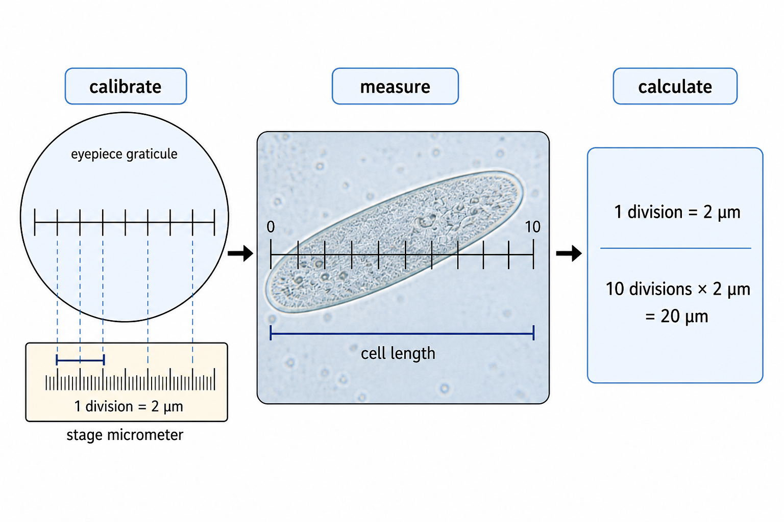
Microscope questions are usually workflow plus maths. Use light microscopes to prepare mounts, stain specimens, and observe cells: first prepare a mount, stain if needed, and focus the specimen. Then calibrate the eyepiece graticule with a stage micrometer so each eyepiece division has a real size. After that, use the triangle: magnification = image size / actual size. Keep units consistent before calculating.
Choose The Right Microscope
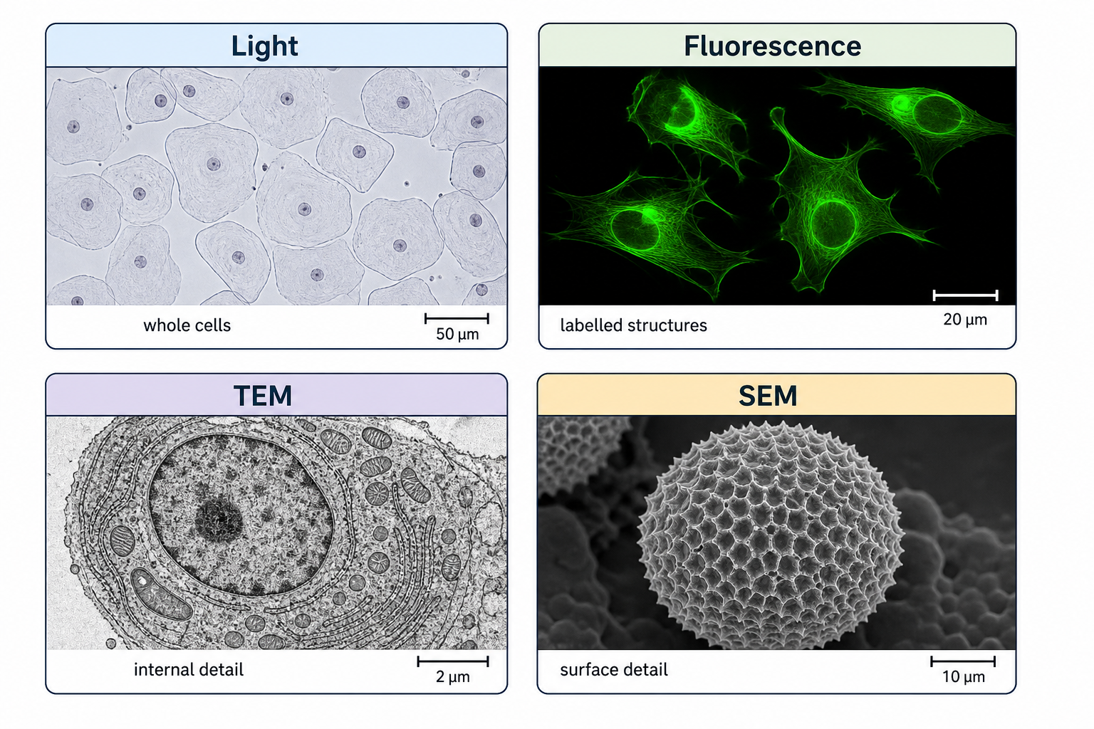
Microscopes should be compared by resolution, not only magnification. Resolution decides whether detail can actually be separated. Use TEM for internal ultrastructure in thin sections, SEM for surface detail, cryogenic EM for fine near-native molecular detail, fluorescence to locate fluorescent molecules, and immunofluorescence when antibodies are used to label specific proteins or structures.
Compare what each method reveals instead of ranking them by magnification alone.
Match the method to the evidence it is best for.
MatchMatch the method to the evidence it is best for.
ChooseFind The Four Universal Cell Parts
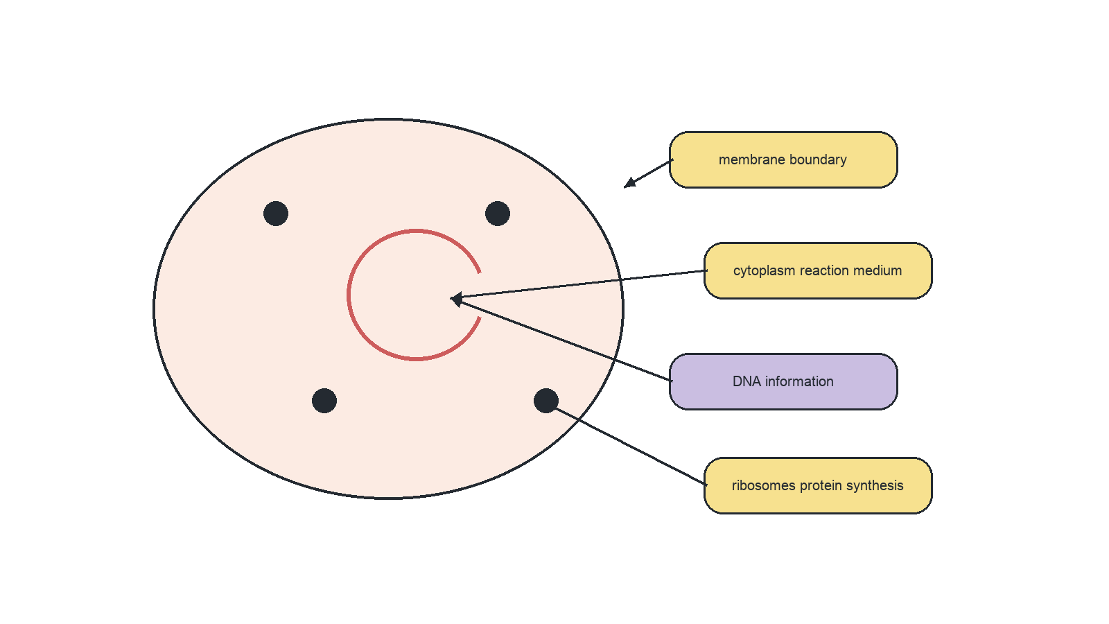
Every cell, prokaryotic or eukaryotic, needs the same four essentials: a plasma membrane, cytoplasm, DNA, and ribosomes. The membrane controls exchange, cytoplasm is the site of many metabolic reactions, DNA stores genetic information, and ribosomes make proteins. These are universal; membrane-bound organelles are not.
The same four essentials appear in every cell type.
Match each universal part to its function.
MatchMatch each universal part to its function.
ChooseDiagnose A Prokaryote
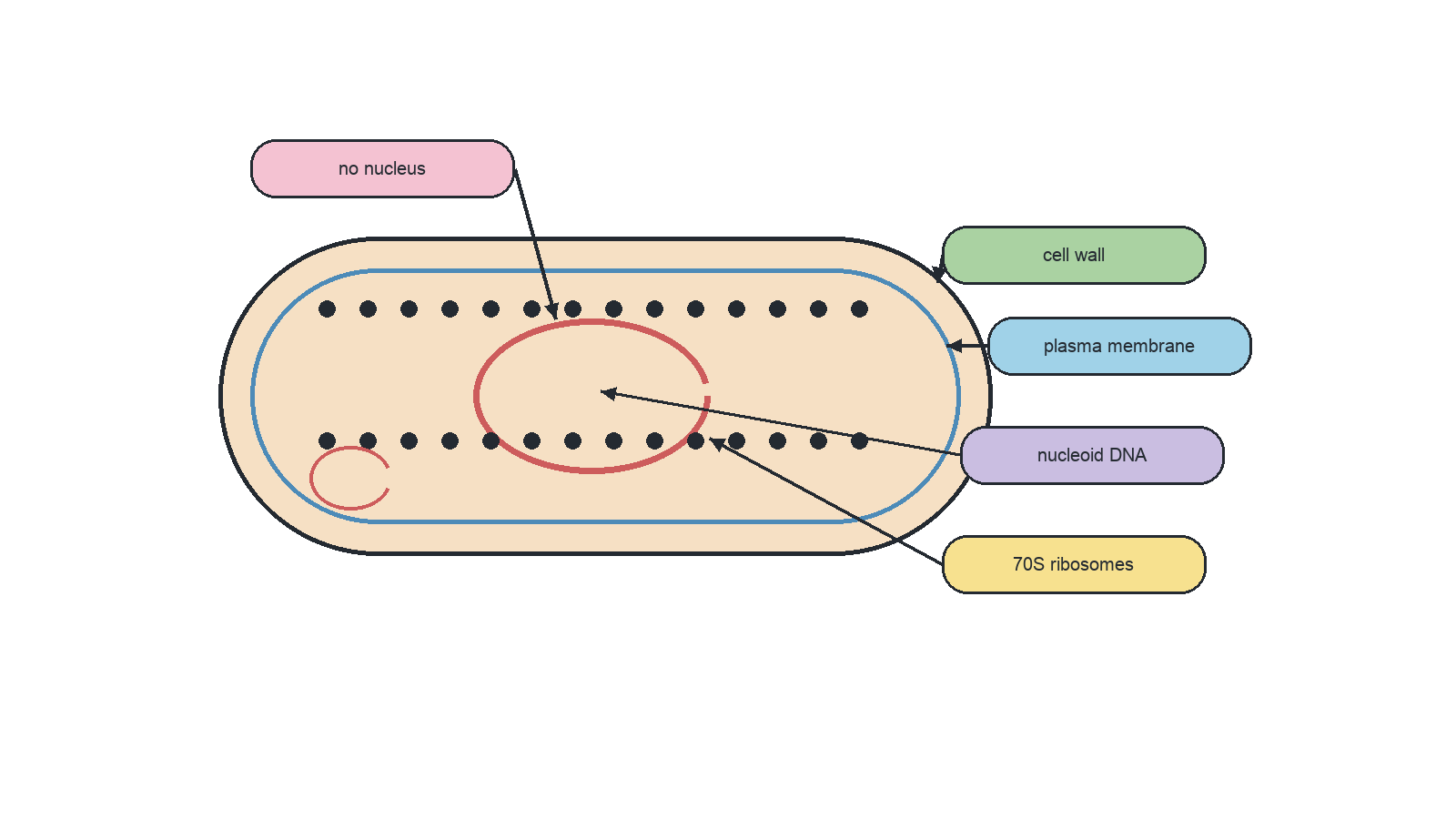
To identify a prokaryote, look first for what is missing: no nucleus and no membrane-bound organelles. Then look for the positive evidence: cell wall, plasma membrane, cytoplasm, 70S ribosomes, naked circular DNA, and sometimes plasmids. E. coli, Bacillus, and Staphylococcus are bacterial examples.
Diagnose prokaryotes by what they lack and by how their DNA is arranged.
Place the labels that make this a bacterial cell.
LabelPlace the labels that make this a bacterial cell.
ChooseMap Compartmentalized Eukaryotic Cells
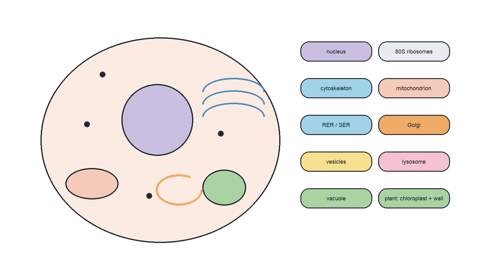
Eukaryotic cells are built around compartmentalization. They have a nucleus, 80S ribosomes, cytoskeleton, and organelles such as mitochondria, ER, Golgi apparatus, vesicles, lysosomes, and vacuoles. Plant cells may add chloroplasts, a large permanent vacuole, and a cellulose cell wall. The key idea is that compartments allow different functions in different spaces.
A eukaryotic cell is defined by compartmentalized cytoplasm around a nucleus.
Sort each structure by where it belongs.
SortSort each structure by where it belongs.
ChooseShow One Cell Doing Life
Unicellular organisms are the strongest proof that one cell can be a whole organism. Amoeba, Chlamydomonas, and Escherichia coli each carry out life processes in one cell: nutrition, metabolism, response, excretion, homeostasis, growth, and reproduction. The examples matter because they turn cell theory from a definition into evidence.
Match each unicellular example to a process it can show.
MatchCompare Plant, Animal, And Fungal Cells
Practice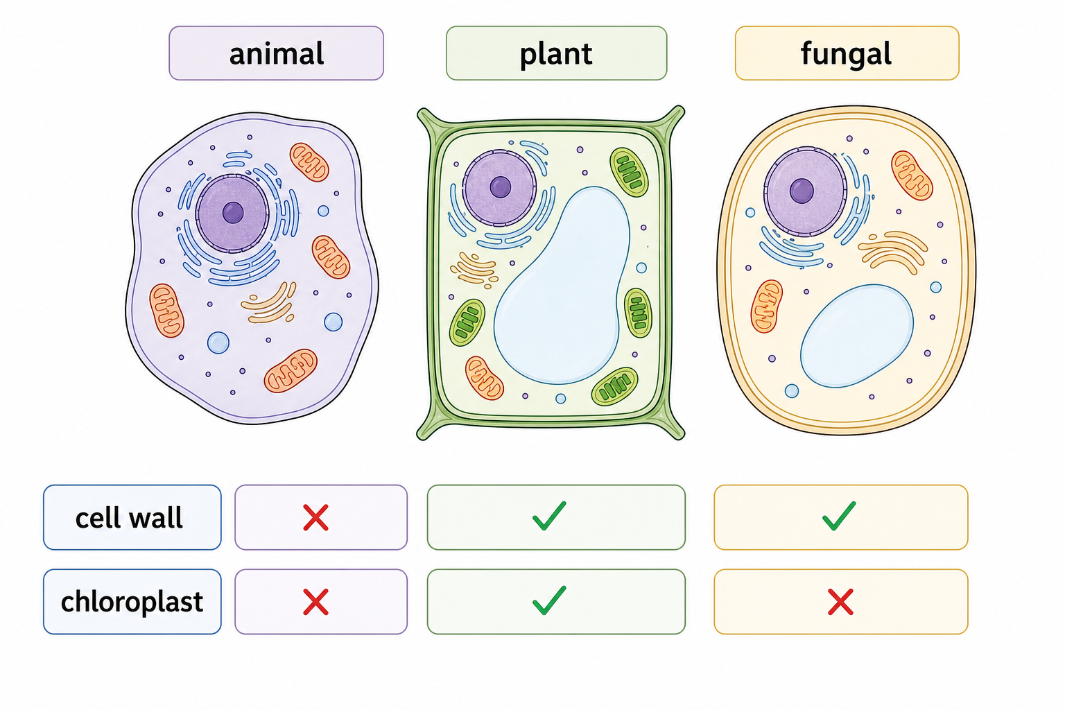
Eukaryotic cell types are best compared feature by feature. Plant cells have cellulose walls, chloroplasts, and large permanent vacuoles. Fungal cells have a cell wall but no chloroplasts. Animal cells lack a cell wall and may have centrioles, cilia, flagella, lysosomes, and temporary vacuoles.
Sort each feature into the best cell-type category.
SortHandle Atypical Cells Without Panic
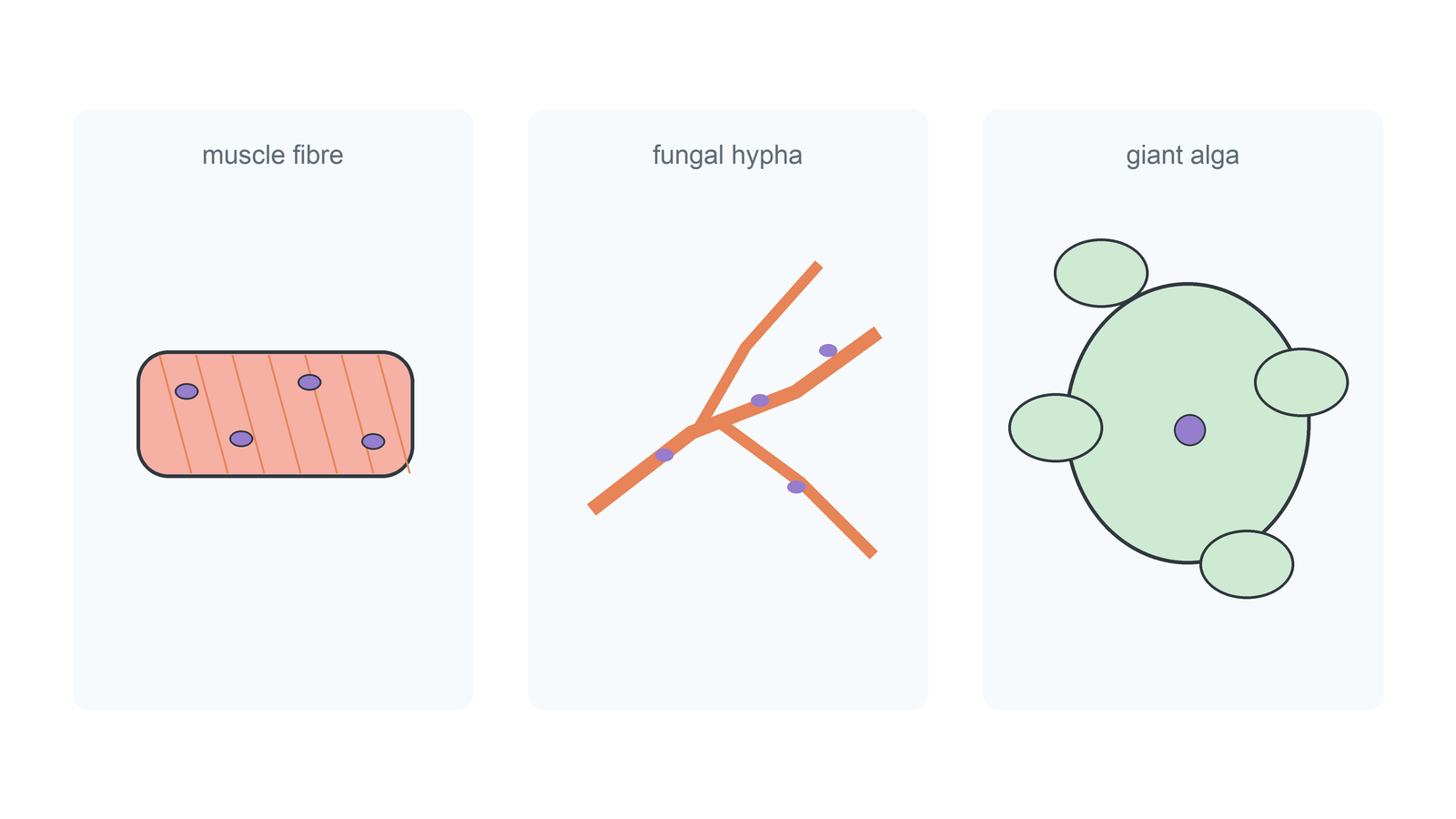
Atypical cells show that textbook diagrams are simplified models. Multinucleate fungal hyphae and striated muscle fibres do not fit the “one cell, one nucleus” picture. Red blood cells and phloem sieve tubes lack nuclei at maturity because of specialization. These cases limit simple cell theory wording, but they do not make the structures non-living or irrelevant.
Fix the student claim.
Spot ErrorsIdentify A Cell From Evidence
PracticeMicrograph identification is evidence work. First name visible structures: nucleus, chloroplasts, mitochondria, ER, Golgi, vacuoles, ribosomes, or cell wall. Then use scale and context. A good answer sounds like “This is likely a plant cell because a cell wall, chloroplasts, and large vacuole are visible,” not “I think it looks plant-like.”
Which justification is strongest for identifying a plant cell in a micrograph?
ChooseDraw Only What The Micrograph Shows
Practice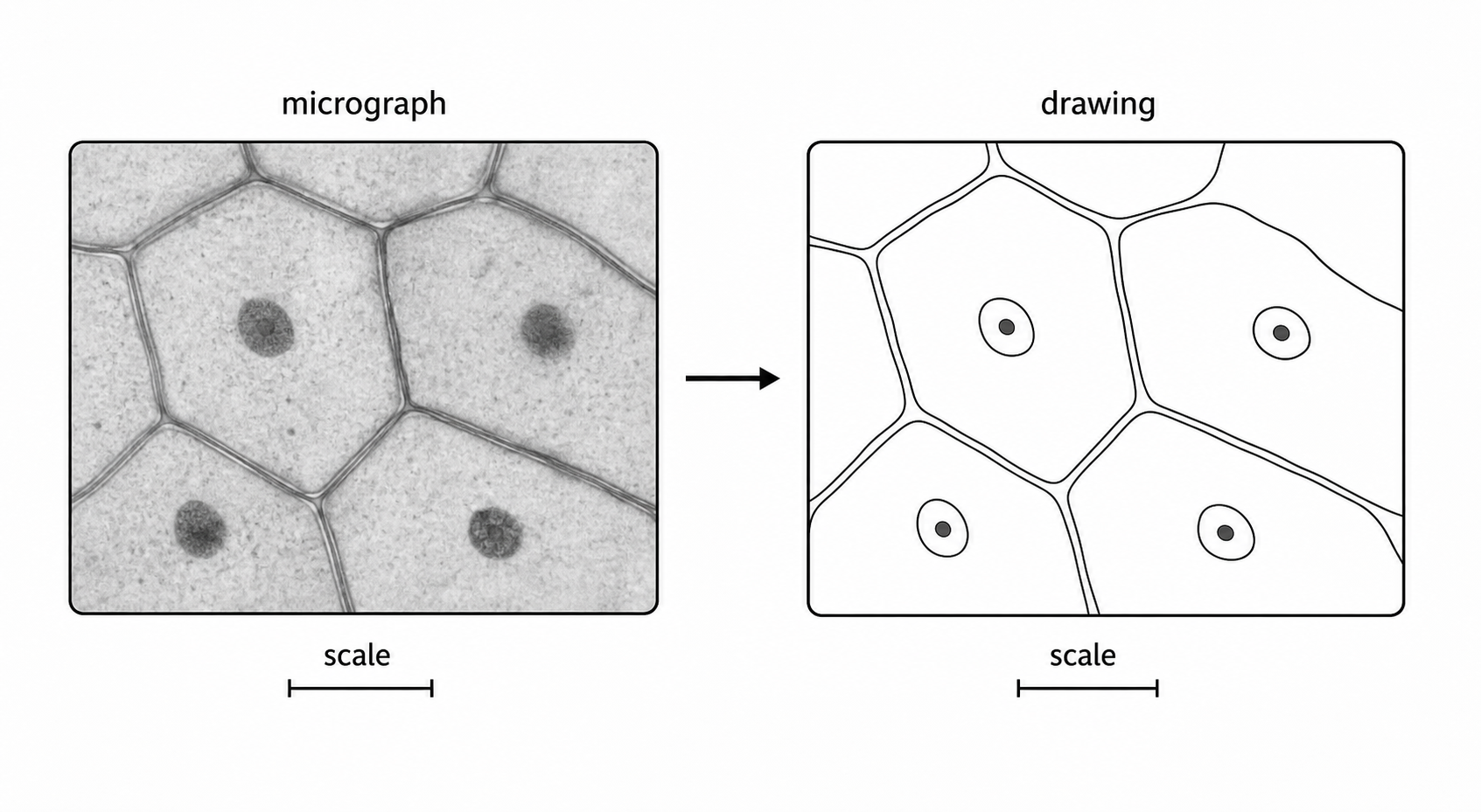
Biological drawing is controlled observation. Draw clear outlines from the electron micrograph, avoid shading, label only visible organelles, and include magnification or scale information. If the question asks for annotations, link each function to the structure you actually drew. The drawing should communicate evidence, not artistic detail. Annotate structures with functions where required, but only when the function belongs to a visible labelled structure.
Spot the mark-losing choices in this drawing plan.
Spot ErrorsSL Retrieval: Read, Identify, Draw
Exam PracticeThe SL core is a practical chain. First, understand cells as structural and functional units. Then use microscopes correctly: prepare, stain, calibrate, measure, and choose a method based on resolution and the detail needed. Finally, identify cell types from visible evidence and draw only what the micrograph shows. This is how the topic turns from definitions into exam performance.
Match each SL task to the rule that saves marks.
MatchUse this for combined SL questions involving cell identification, microscopy, or biological drawing.
Use this for combined SL questions involving cell identification, microscopy, or biological drawing.
A strong SL answer uses evidence from the image and the scale. For example, a prokaryotic cell is identified by small scale, no nucleus, naked circular DNA or simple internal structure, whereas a plant cell is supported by visible cell wall, chloroplasts, and a large vacuole. Calculations require calibration and unit conversion, and drawings should use clear outlines, labels, and scale information without shading or invented structures.
Listing organelles from memory without using the micrograph evidence.