
Read The Amino Acid Backbone
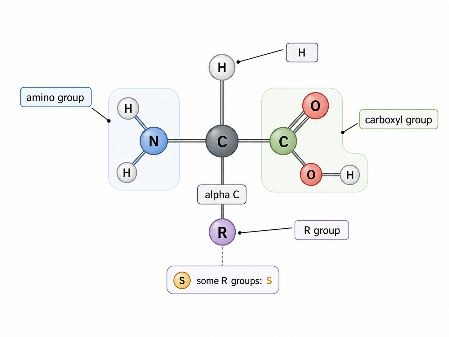
Every amino acid has the same backbone: an alpha carbon bonded to an amine group, a carboxyl group, a hydrogen atom, and an R-group. The R-group is the variable part, so it determines properties such as charge, polarity, hydrophobicity, and later folding behaviour. Proteins contain C, H, O, N, and usually S because some amino acids contain sulfur.
Use the shared backbone to recognize any amino acid, then look to the R-group for its chemistry.
Label the generalized amino acid.
LabelLabel the generalized amino acid.
ChooseBuild A Peptide Bond
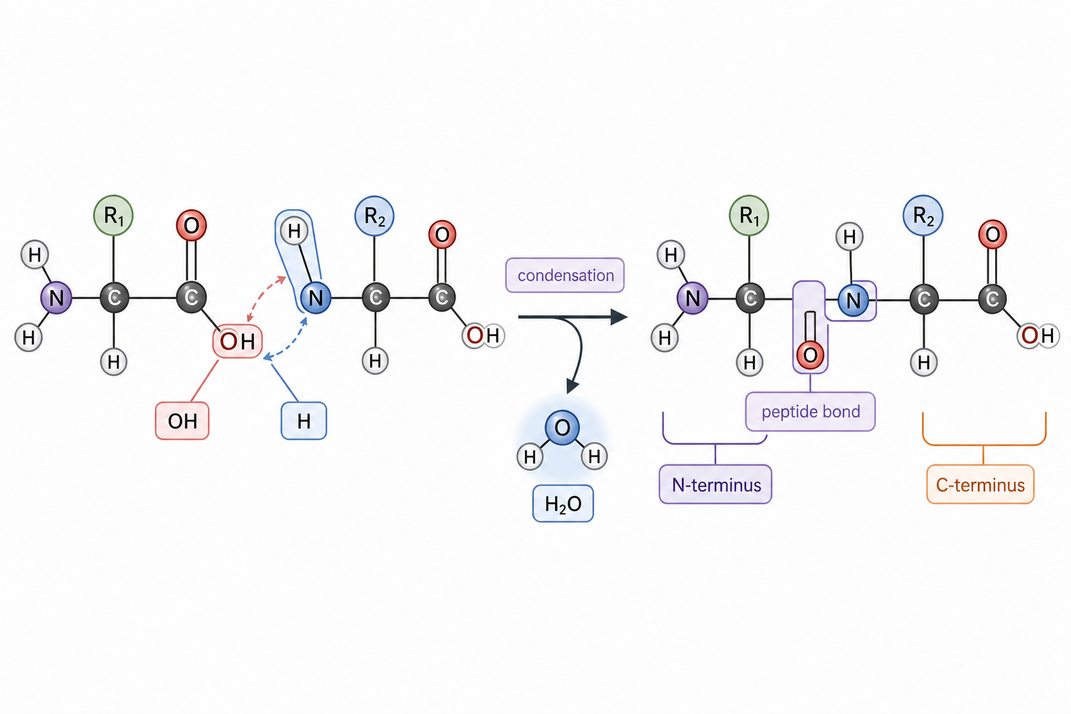
Peptide bonds form by condensation. The carboxyl group of one amino acid reacts with the amine group of another, water is released, and a peptide bond links the residues. The chain has direction: an N-terminus at one end and a C-terminus at the other. Ribosomes assemble polypeptides in this directional order.
Track which atoms leave as water, then identify the new peptide bond and chain direction.
Put peptide-bond formation in order.
OrderPut peptide-bond formation in order.
ChooseDecide Which Amino Acids Must Come From Diet
PracticeEssential amino acids are “essential” because the body cannot synthesize enough of them, so they must come from dietary protein. Non-essential amino acids can be made by transamination, mainly in the liver. If an essential amino acid is missing, protein synthesis is limited because the ribosome cannot complete all needed polypeptides, which can contribute to malnutrition.
Sort each statement into essential, non-essential, or deficiency consequence.
SortExplain Protein Sequence Diversity
Protein diversity comes from sequence possibilities. Twenty coded amino acids can be combined in different types, numbers, and orders, creating vast numbers of polypeptide sequences. Genes encode those sequences, and the proteome is the full set of proteins expressed by a cell, tissue, or organism at a given time.
Match each term to its role in protein diversity.
MatchPredict Denaturation From Shape Change
Practice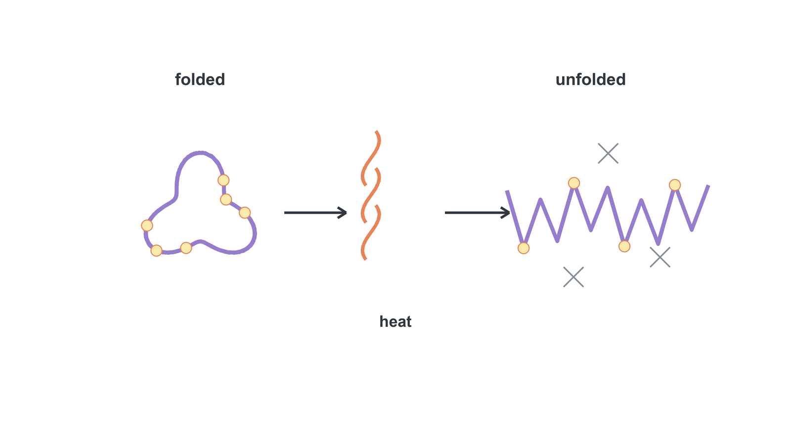
Protein shape determines function, especially for enzyme active sites. High temperature or unsuitable pH can disrupt weak bonds that maintain the folded shape. When the active site changes shape, the substrate no longer fits properly and function falls. Small proteins may sometimes refold, but denaturation is often irreversible.
An enzyme is moved to a very acidic solution. Predict what happens.
PredictHL: Classify R-Group Chemistry
R-groups are the chemical personalities of amino acids. They may be acidic, basic, polar hydrophilic, or non-polar hydrophobic. This chemistry determines solubility, interactions, folding, and function. In soluble proteins, hydrophobic R-groups are often buried away from water, while polar or charged groups are more likely to face water.
Sort each R-group description by chemistry and folding effect.
SortHL: Trace DNA To Primary Structure
Primary structure is the ordered amino acid sequence joined by peptide bonds. DNA controls that sequence through mRNA during translation. Because the sequence determines which R-groups appear and where, even one amino acid substitution can change interactions, conformation, and protein properties.
Match each step from gene to protein property.
MatchHL: Inspect Alpha Helices And Beta Sheets
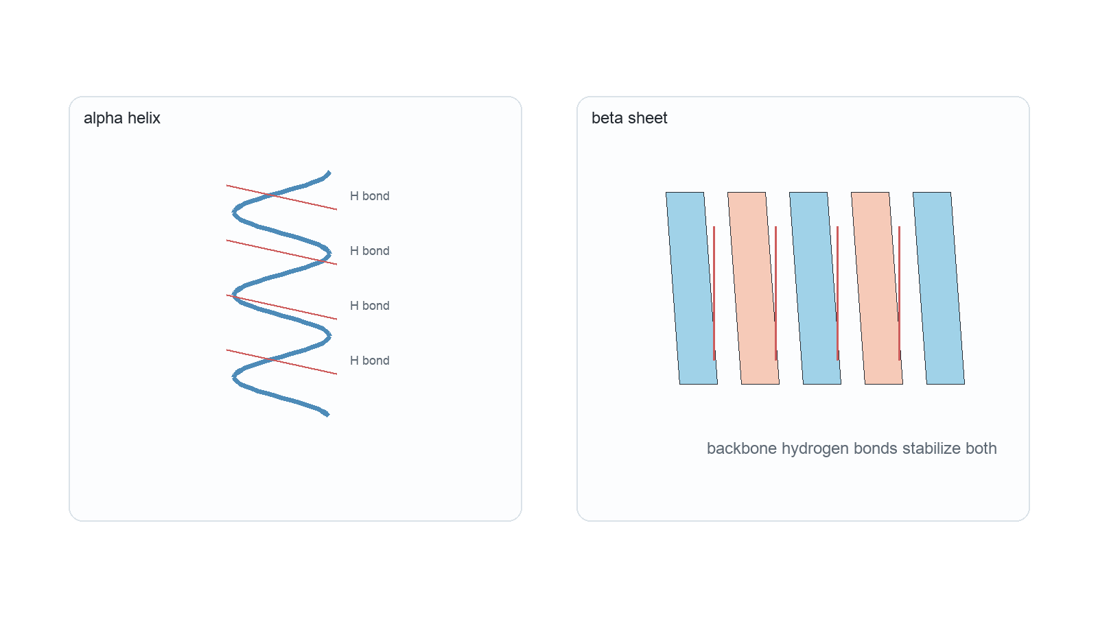
Secondary structure is local folding of the polypeptide backbone. Some regions coil into alpha helices; others pleat into beta-sheets. Both are stabilized by regular hydrogen bonding between backbone groups, not mainly by R-group interactions. These motifs can combine into domains such as coiled coils or beta sandwiches.
Both patterns come from regular hydrogen bonding, but one coils and the other pleats.
Match each secondary-structure term to its meaning.
MatchMatch each secondary-structure term to its meaning.
ChooseHL: Stabilize Tertiary Folding
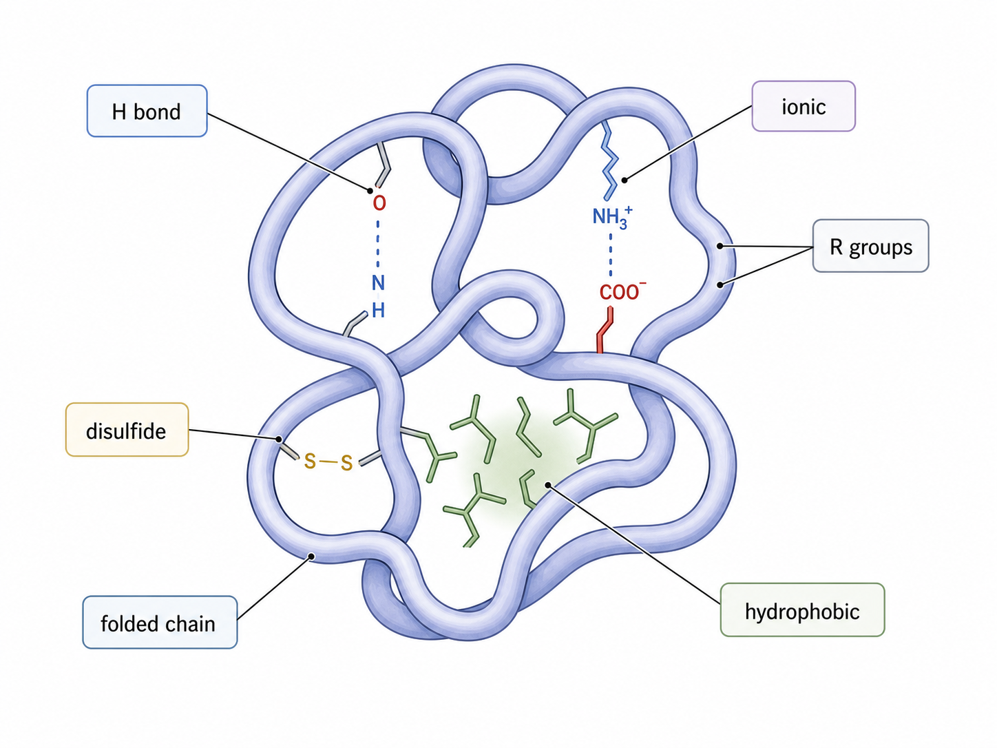
Tertiary structure is the unique 3D folding of one polypeptide chain. It is stabilized by interactions between R-groups: hydrogen bonds, ionic bonds, disulfide covalent bonds between cysteine residues, and hydrophobic interactions that cluster non-polar groups away from water. This folding creates the final shape needed for function.
Different side-chain interactions all help stabilize one functional 3D shape.
Match each tertiary interaction to its source.
MatchMatch each tertiary interaction to its source.
ChooseHL: Place Residues In Soluble And Membrane Proteins
Practice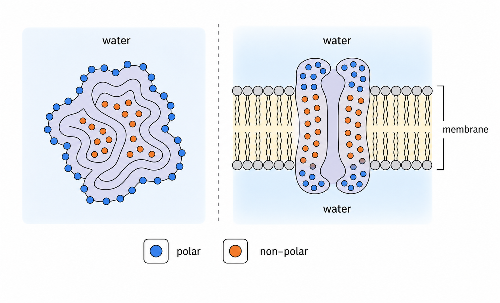
Residue placement depends on environment. In soluble globular proteins, hydrophobic residues are often buried in the core, while polar and charged residues are commonly exposed to water. Integral membrane proteins reverse part of that logic: hydrophobic regions face lipid tails in the bilayer, while hydrophilic regions face watery environments.
Compare residue placement in a soluble protein versus a membrane protein.
CompareHL: Compare Quaternary Protein Examples
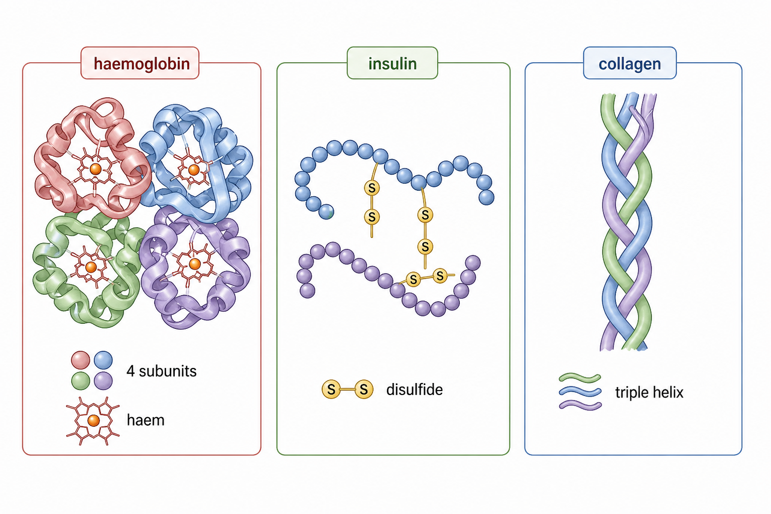
Quaternary structure means two or more polypeptide chains join into one functional protein. Haemoglobin is conjugated because it has four globin chains plus non-polypeptide haem groups with iron. Insulin and collagen are non-conjugated examples: insulin is stabilized by disulfide bonds, while collagen forms a triple helix.
All three are quaternary examples, but they are built and stabilized in different ways.
Match each protein example to its structural clue.
MatchMatch each protein example to its structural clue.
ChooseHL: Distinguish Globular And Fibrous Proteins
PracticeGlobular and fibrous proteins differ in shape, solubility, and job. Globular proteins are compact, often soluble, and suited to transport, signalling, or catalysis; insulin is a small globular hormone stabilized by disulfide bridges. Fibrous proteins are long, insoluble, and structural; collagen provides tensile strength.
Sort each feature into globular or fibrous protein logic.
SortCore Transfer: Build And Use Proteins
Exam PracticeThe core protein story is build -> vary -> function. Amino acids share a backbone but differ in R-groups. Peptide bonds form by condensation between carboxyl and amine groups. Some amino acids must come from diet, or protein synthesis is limited. Twenty coded amino acids create many sequences by type, number, and order. Finally, shape determines function, so denaturation changes performance.
Match each core prompt to its answer rule.
MatchUse this for core protein questions on amino acid structure, peptide-bond formation, essential amino acids, sequence diversity, or denaturation.
Use this for core protein questions on amino acid structure, peptide-bond formation, essential amino acids, sequence diversity, or denaturation.
Proteins are built from amino acids with a shared alpha-carbon backbone and variable R-groups that determine chemical properties. Peptide bonds form by condensation between the carboxyl group of one amino acid and the amine group of another, releasing water and creating directional chains. Essential amino acids must be obtained from dietary protein, while non-essential amino acids can be made by transamination. Protein diversity comes from the type, number and order of the 20 coded amino acids. Protein shape determines function, so high temperature or unsuitable pH can disrupt weak bonds, denature the protein and reduce activity.
Listing terms without explaining how structure leads to function.
HL Transfer: Explain Folding Levels
Exam PracticeHL protein questions are level-control questions. R-group chemistry predicts solubility and interactions. Primary structure is the DNA-coded amino acid sequence. Secondary structure is local alpha helix or beta-sheet stabilized by backbone hydrogen bonds. Tertiary structure is one polypeptide’s 3D fold stabilized by R-group interactions. Quaternary structure joins multiple chains. Examples such as haemoglobin, insulin, and collagen anchor these levels in real proteins.
Match each HL clue to the correct protein-structure level or example.
MatchUse this for HL questions on R-group chemistry, protein-structure levels, residue placement, and named examples.
Use this for HL questions on R-group chemistry, protein-structure levels, residue placement, and named examples.
R-group chemistry determines solubility, interactions, folding and function. Primary structure is the amino acid sequence controlled by DNA via mRNA; a single amino acid change can alter conformation. Secondary structures such as alpha helices and beta-sheets are stabilized by backbone hydrogen bonds. Tertiary structure is the unique 3D fold of one polypeptide, stabilized by R-group hydrogen bonds, ionic bonds, disulfide bonds and hydrophobic interactions. Quaternary structure joins two or more polypeptide chains, as in haemoglobin with four globin chains and haem groups. Soluble globular proteins often bury hydrophobic residues, while membrane proteins expose hydrophobic regions to lipid tails. Insulin is globular; collagen is fibrous and gives tensile strength.
Mixing up structure levels or giving named examples without structural reasons.