EduNinja
Question rangeA2.2.3—Developments in microscopy
[Maximum number: 1]

The images of the radiolarian, a single-celled marine organism, were produced using a light microscope (left) and a scanning electron microscope (right).

Question image

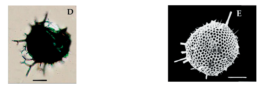
What is a reason for the difference in quality of these images?

A

Light cannot pass through the specimen.

B

Higher magnification can be achieved with the electron microscope.

C

The resolution of the electron microscope is higher.

D

Samples are stained with methylene blue when viewed with the light microscope.

[Maximum number: 5]

The image below shows freely-growing aerial rhizoids of the bryophyte Homalothecium sericeum near a shoot apex.
Removed for copyright reasons

(a)

Calculate the magnification of this image.

[ 1 ]
(b)

Outline the procedure for focusing a light microscope.

[ 3 ]
(c)

State one advantage of using a light microscope compared to an electron microscope.

[ 1 ]
[Maximum number: 4]

The figure shows a transmission electron micrograph of rotavirus particles. Each rotavirus is about 70 nanometres in diameter.

Question image
(a)

State a reason for using an electron microscope to view this virus rather than a light microscope.

[ 1 ]
(b)

Rotavirus causes diarrhea and vomiting. Explain why viral diseases cannot be treated using antibiotics.

[ 2 ]
(c)

State an application of plasmids in biotechnology.

[ 1 ]
[Maximum number: 1]

What is a feature of immunofluorescence in light microscopy?

A

Can only be used on dead cells

B

Enables higher resolution

C

Attaches a fluorescent stain to an antibody

D

Attaches a fluorescent stain to an antigen

0